
Page 705 - Veterinary Immunology, 10th Edition
P. 705
Peyer's patches are secondary lymphoid organs (see Fig. 12.6). In
VetBooks.ir lambs, the ileocecal patches increase in size from birth to 6 months
of age and then regress, leaving only a small scar. In contrast, the
jejunal patches persist throughout adult life and continue to play a
major role in intestinal defense.
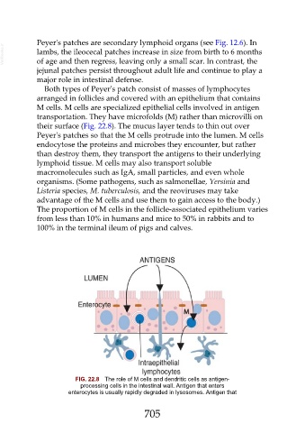
Both types of Peyer's patch consist of masses of lymphocytes
arranged in follicles and covered with an epithelium that contains
M cells. M cells are specialized epithelial cells involved in antigen
transportation. They have microfolds (M) rather than microvilli on
their surface (Fig. 22.8). The mucus layer tends to thin out over
Peyer's patches so that the M cells protrude into the lumen. M cells
endocytose the proteins and microbes they encounter, but rather
than destroy them, they transport the antigens to their underlying
lymphoid tissue. M cells may also transport soluble
macromolecules such as IgA, small particles, and even whole
organisms. (Some pathogens, such as salmonellae, Yersinia and
Listeria species, M. tuberculosis, and the reoviruses may take
advantage of the M cells and use them to gain access to the body.)
The proportion of M cells in the follicle-associated epithelium varies
from less than 10% in humans and mice to 50% in rabbits and to
100% in the terminal ileum of pigs and calves.
FIG. 22.8 The role of M cells and dendritic cells as antigen-
processing cells in the intestinal wall. Antigen that enters
enterocytes is usually rapidly degraded in lysosomes. Antigen that
705