
Page 709 - Veterinary Immunology, 10th Edition
P. 709
VetBooks.ir
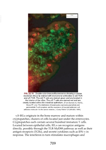
FIG. 22.10 Double-color immunofluorescence showing a canine
duodenal villous tip stained with monoclonal antibodies to α/β TCR
and γ/δ TCR. The α/β T cells are stained green and are located in
the interior of the villus. The γ/δ T cells are stained red and are
clearly located within the intestinal epithelium. (From German AJ, Hall EJ,
Moore PF, et al: The distribution of lymphocytes expressing alpha/beta and
gamma/delta T-cell receptors, and the expression of mucosal addressin cell
adhesion molecule-1 in the canine intestine, J Comp Pathol 121:249-263, 1999.)
γ/δ IELs originate in the bone marrow and mature within
cryptopatches, clusters of cells located just under the enterocytes.
Cryptopatches each contain several hundred immature T cells.
Located between epithelial cells, IELs can recognize antigens
directly, possibly through the TLR-MyD88 pathway as well as their
antigen receptors (TCRs), and secrete cytokines such as IFN-γ in
response. The interferon in turn stimulates macrophages and
709