
Page 714 - Veterinary Immunology, 10th Edition
P. 714
IgA monomers are about 160 kDa in size and are typical four-
VetBooks.ir chain, Y-shaped molecules (see Fig. 16.6). They are usually secreted
as dimers or larger polymers linked by a J chain. IgA has several
extra cysteine residues in its heavy chains. As a result, the short
interchain disulfide bonds compact the chains and shield
vulnerable bonds from proteases.
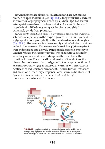
IgA is synthesized and secreted by plasma cells in the intestinal
submucosa, especially in the crypt region. This dimeric IgA binds to
a glycoprotein receptor (pIgR) on the basal surface of enterocytes
(Fig. 22.13). The receptor binds covalently to the Cα2 domain of one
of the IgA monomers. The membrane-bound IgA-pIgR complex is
then endocytosed and actively transported across the enterocyte.
When it reaches the exterior surface, this endocytic vesicle fuses
with the plasma membrane and exposes the complex to the
intestinal lumen. The extracellular domains of the pIgR are then
cleaved by proteases so that the IgA, with the receptor peptide still
attached (secretory IgA), is released into the lumen. The receptor
peptide is called secretory component. The production, transport,
and secretion of secretory component occur even in the absence of
IgA so that free secretory component is found in high
concentrations in intestinal contents.
FIG. 22.13 IgA is secreted by mucosal plasma cells and binds to
receptors (pIgR) on the interior surface of intestinal enterocytes.
The bound IgA is taken into the enterocytes and passed in vesicles
714