
Page 368 - The Toxicology of Fishes
P. 368
348 The Toxicology of Fishes
Ha
Hv
Rdc Sv
Ldc
LD
L HV L
Myv
GB GB
GB
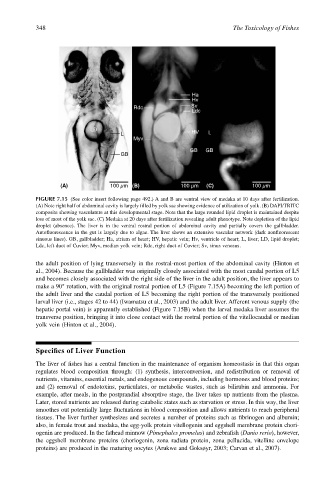
(A) 100 µm (B) 100 µm (C) 100 µm
FIGURE 7.15 (See color insert following page 492.) A and B are ventral view of medaka at 10 days after fertilization.
(A) Note right half of abdominal cavity is largely filled by yolk sac showing evidence of utilization of yolk. (B) DAPI/TRITC
composite showing vasculature at this developmental stage. Note that the large rounded lipid droplet is maintained despite
loss of most of the yolk sac. (C) Medaka at 20 days after fertilization revealing adult phenotype. Note depletion of the lipid
droplet (absence). The liver is in the ventral rostral portion of abdominal cavity and partially covers the gallbladder.
Autofluorescence in the gut is largely due to algae. The liver shows an extensive vascular network (dark nonfluorescent
sinuous lines). GB, gallbladder; Ha, atrium of heart; HV, hepatic vein; Hv, ventricle of heart; L, liver; LD, lipid droplet;
Ldc, left duct of Cuvier; Myv, median yolk vein; Rdc, right duct of Cuvier; Sv, sinus venosus.
the adult position of lying transversely in the rostral-most portion of the abdominal cavity (Hinton et
al., 2004). Because the gallbladder was originally closely associated with the most caudal portion of L5
and becomes closely associated with the right side of the liver in the adult position, the liver appears to
make a 90° rotation, with the original rostral portion of L5 (Figure 7.15A) becoming the left portion of
the adult liver and the caudal portion of L5 becoming the right portion of the transversely positioned
larval liver (i.e., stages 42 to 44) (Iwamatsu et al., 2003) and the adult liver. Afferent venous supply (the
hepatic portal vein) is apparently established (Figure 7.15B) when the larval medaka liver assumes the
transverse position, bringing it into close contact with the rostral portion of the vitellocaudal or median
yolk vein (Hinton et al., 2004).
Specifics of Liver Function
The liver of fishes has a central function in the maintenance of organism homeostasis in that this organ
regulates blood composition through: (1) synthesis, interconversion, and redistribution or removal of
nutrients, vitamins, essential metals, and endogenous compounds, including hormones and blood proteins;
and (2) removal of endotoxins, particulates, or metabolic wastes, such as bilirubin and ammonia. For
example, after meals, in the postprandial absorptive stage, the liver takes up nutrients from the plasma.
Later, stored nutrients are released during catabolic states such as starvation or stress. In this way, the liver
smoothes out potentially large fluctuations in blood composition and allows nutrients to reach peripheral
tissues. The liver further synthesizes and secretes a number of proteins such as fibrinogen and albumin;
also, in female trout and medaka, the egg-yolk protein vitellogenin and eggshell membrane protein chori-
ogenin are produced. In the fathead minnow (Pimephales promelas) and zebrafish (Danio rerio), however,
the eggshell membrane proteins (choriogenin, zona radiata protein, zona pellucida, vitelline envelope
proteins) are produced in the maturing oocytes (Arukwe and Goksøyr, 2003; Carvan et al., 2007).