
Page 161 - Veterinary Histology of Domestic Mammals and Birds, 5th Edition
P. 161
Blood and haemopoiesis (sanguis et haemocytopoesis) 143
VetBooks.ir rER · appearance of azurophilic granules,
· alteration in the shape of the nucleus and
· decondensation of chromatin.
MONOCYTE MORPHOLOGY AND FUNCTION
Monocytes are classified as agranulocytes. In their mature
form, these mononuclear cells are the largest leucocytes
Golgi in the blood (12–20 μm) and represent 2–10% of circulat-
apparatus ing white blood cells. The shape of the nucleus is variable
(often round, sometimes kidney-shaped or amoeboid)
rER and the abundant cytoplasm is weakly basophilic with
numerous mitochondria and clusters of Golgi cisternae.
There are few ribosomes and the rER is poorly developed.
Lymph Monocytes may contain azurophilic granules. These
are lysosomes and thus contain numerous proteolytic
enzymes. On the cell surface there are irregularly formed
finger-like structures (pseudopodia) or individual micro-
7.11 Fine structure of an active plasma cell in the villi that contribute to motility and phagocytic activity.
medulla of a lymph node (x8000). Monocytes circulate (as ‘blood macrophages’) for a short
period (2 days) before actively departing the blood vascu-
Monocytes lar system and passing via amoeboid movement through
Monocytes have numerous functions, particularly in innate the interstitial tissue. Their lifespan, once in the tissues, is
immunity and immunoregulation. unclear but is estimated at 60–90 days.
After leaving the blood circulation, monocytes trans-
DEVELOPMENT OF MONOCYTES form into tissue macrophages. These include specific cell
(MONOCYTOPOIESIS) types such as Kupffer cells (liver), alveolar macrophages
Division and differentiation of haemopoietic stem cells and (lung), peritoneal macrophages, sinus endothelial cells
progenitor cells in the bone marrow initially gives rise to (lymphoid organs), histiocytes (connective tissue) and
monoblasts (Figure 7.12). These develop further into pro- osteoclasts. Macrophages are an important component of
monocytes (diameter 16–22 μm) containing azurophilic the mononuclear phagocyte system, sometimes referred
granules. After frequent mitotic divisions over the course to as the reticuloendothelial system.
of a few days, mature monocytes are formed (Figure Through their ability to phagocytose antigenic mate-
7.12). It appears that the early stages of development are rial, monocytes are involved in innate immune responses.
shared with those of neutrophils, and that monocytes may They secrete complement and synthesise interferon.
arise from promyelocytes. The phagocytic potential of Monocytes also contribute to the destruction of aged red
both cells may thus reflect a common origin. blood cells in the spleen, and to iron and fat metabolism.
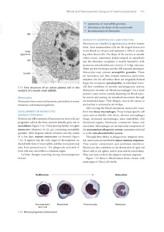
Cellular changes occurring during monocytopoiesis They also have a role in the adaptive immune response.
include: Figure 7.13 shows a blood smear from a horse with
many types of blood cell visible.
7.12 Monocytopoiesis (schematic).
Vet Histology.indb 143 16/07/2019 14:58