
Page 158 - Veterinary Histology of Domestic Mammals and Birds, 5th Edition
P. 158
140 Veterinary Histology of Domestic Mammals and Birds
pigs. Eosinophils form 2–10% of circulating leucocytes, a Agranulocytes
VetBooks.ir considerably lower proportion than neutrophils. Normal Agranulocytes lack distinctive granules. These cells
values vary with species (Table 7.2; chickens 2%).
include:
Eosinophils measure 12–14 μm in diameter and have
a segmented nucleus. They exhibit amoeboid motility and · lymphocytes and
phagocytic activity, the latter being oriented particularly · monocytes.
towards antigen–antibody complexes. Thus, an increase
in circulating eosinophils (eosinophilia) is commonly Lymphocytes
observed in association with allergic responses (allergies, Lymphocytes are the most numerous agranulocytes
parasitism). (Figures 7.8 to 7.10). Only around 2% of these round,
In these instances, eosinophils are activated by the basophilic cells circulate in the blood, the majority being
release of chemical mediators (leukotrienes, histamine concentrated in lymphoid organs (for further detail see
released by mast cells). Eosinophils also produce substances Chapter 8, ‘Immune system and lymphatic organs’).
that inhibit the release of histamine, thus down-regulating The proportion of lymphocytes in the circulating leu-
the inflammatory response. Adrenocorticotropic (ACTH) cocyte population varies with species (cattle, sheep, goats:
hormone and cortisol have a negative effect on circulating 50–70%; horses and cats: 30–35%; dogs: 20–25%) (Table
eosinophil numbers, potentially causing eosinopenia. 7.2).
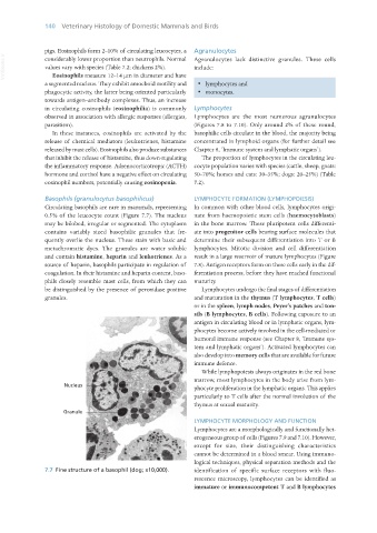
Basophils (granulocytus basophilicus) LYMPHOCYTE FORMATION (LYMPHOPOIESIS)
Circulating basophils are rare in mammals, representing In common with other blood cells, lymphocytes origi-
0.5% of the leucocyte count (Figure 7.7). The nucleus nate from haemopoietic stem cells (haemocytoblasts)
may be bilobed, irregular or segmented. The cytoplasm in the bone marrow. These pluripotent cells differenti-
contains variably sized basophilic granules that fre- ate into progenitor cells bearing surface molecules that
quently overlie the nucleus. These stain with basic and determine their subsequent differentiation into T or B
metachromatic dyes. The granules are water soluble lymphocytes. Mitotic division and cell differentiation
and contain histamine, heparin and leukotrienes. As a result in a large reservoir of mature lymphocytes (Figure
source of heparin, basophils participate in regulation of 7.8). Antigen receptors form on these cells early in the dif-
coagulation. In their histamine and heparin content, baso- ferentiation process, before they have reached functional
phils closely resemble mast cells, from which they can maturity.
be distinguished by the presence of peroxidase positive Lymphocytes undergo the final stages of differentiation
granules. and maturation in the thymus (T lymphocytes, T cells)
or in the spleen, lymph nodes, Peyer’s patches and ton-
sils (B lymphocytes, B cells). Following exposure to an
antigen in circulating blood or in lymphatic organs, lym-
phocytes become actively involved in the cell-mediated or
humoral immune response (see Chapter 8, ‘Immune sys-
tem and lymphatic organs’). Activated lymphocytes can
also develop into memory cells that are available for future
immune defence.
While lymphopoiesis always originates in the red bone
marrow, most lymphocytes in the body arise from lym-
phocyte proliferation in the lymphatic organs. This applies
particularly to T cells after the normal involution of the
thymus at sexual maturity.
LYMPHOCYTE MORPHOLOGY AND FUNCTION
Lymphocytes are a morphologically and functionally het-
erogeneous group of cells (Figures 7.9 and 7.10). However,
except for size, their distinguishing characteristics
cannot be determined in a blood smear. Using immuno-
logical techniques, physical separation methods and the
7.7 Fine structure of a basophil (dog; x10,000). identification of specific surface receptors with fluo-
rescence microscopy, lymphocytes can be identified as
immature or immunocompetent T and B lymphocytes
Vet Histology.indb 140 16/07/2019 14:58