
Page 325 - Veterinary Histology of Domestic Mammals and Birds, 5th Edition
P. 325
Female reproductive system (organa genitalia feminina) 307
VetBooks.ir
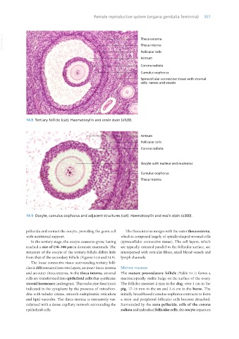
14.8 Tertiary follicle (cat). Haematoxylin and eosin stain (x120).
14.9 Oocyte, cumulus oophorus and adjacent structures (cat). Haematoxylin and eosin stain (x300).
pellucida and contact the oocyte, providing the germ cell The theca interna merges with the outer theca externa,
with nutritional support. which is composed largely of spindle-shaped stromal cells
In the tertiary stage, the oocyte ceases to grow, having (spinocellular connective tissue). The cell layers, which
reached a size of 130–300 μm in domestic mammals. The are typically oriented parallel to the follicular surface, are
structure of the oocyte of the tertiary follicle differs little interspersed with reticular fibres, small blood vessels and
from that of the secondary follicle (Figures 14.8 and 14.9). lymph channels.
The loose connective tissue surrounding tertiary folli-
cles is differentiated into two layers, an inner theca interna matuRe follicles
and an outer theca externa. In the theca interna, stromal The mature preovulatory follicle (Table 14.1) forms a
cells are transformed into epithelioid cells that synthesise macroscopically visible bulge on the surface of the ovary.
steroid hormones (androgens). This endocrine function is The follicles measure 2 mm in the dog, over 1 cm in the
indicated in the cytoplasm by the presence of mitochon- pig, 17–18 mm in the ox and 3–6 cm in the horse. The
dria with tubular cristae, smooth endoplasmic reticulum initially broad-based cumulus oophorus contracts to form
and lipid vacuoles. The theca interna is extensively vas- a stem and peripheral follicular cells become detached.
cularised with a dense capillary network surrounding the Surrounded by the zona pellucida, cells of the corona
epithelioid cells. radiata and individual follicular cells, the oocyte separates
Vet Histology.indb 307 16/07/2019 15:05