
Page 326 - Veterinary Histology of Domestic Mammals and Birds, 5th Edition
P. 326
308 Veterinary Histology of Domestic Mammals and Birds
from the follicular wall and floats freely in the follicular oocyte occurs as a secondary process. Morphologically,
VetBooks.ir fluid. In most domestic mammals, the primary oocyte atretic follicles are characterised by shrinking and conden-
(ovocytus primarius) completes meiosis I prior to ovula-
sation of the nucleus, and hyalinisation of the basal lamina.
tion. In the horse and dog, this occurs after ovulation has
taken place. During the completion of meiosis I, genetic ovulation
information is distributed evenly among the two result- Ovulation involves rupture of the mature follicle and
ing nuclei, while the cytoplasm is unequally apportioned. release of the oocyte. This process is brought about
This gives rise to a secondary oocyte (ovocytus secundar- primarily by hormonally mediated changes in the micro-
ius) and a non-viable polar body (polocytus primarius). vasculature and enzymatic lysis of loose connective tissue
Immediately after completion of the first meiotic division at the predetermined site of follicular rupture (stigma
(reduction division), meiosis II (equatorial division) com- folliculare) (Figure 14.11).
mences in the secondary oocyte. This is completed only Rupture of the follicle wall is preceded by a brief period
after penetration of the oocyte by a spermatozoon within of hyperaemia and subsequent narrowing of the capil-
the uterine tube. This division is also uneven, resulting in laries of the theca interna. Interruption of the blood
a haploid ovum, which is capable of being fertilised, and supply to the stigma is followed by complete degenera-
a small, non-functional polar body. tion of the capillary network. This is accompanied by
enzymatic breakdown (collagenases, proteases) of the
folliculaR atResia tunica albuginea and the theca, and a reduction in the
Most primordial and primary follicles, and many follicles number of follicular cells. Breakdown of the follicle wall
that reach later stages of development, undergo regression is mediated by hormones, primarily luteinising hormone
(follicular atresia). This is a normal, continuously occur- (LH), oestrogens and prostaglandins (PGE and PGF ).
2 2α
ring process. Only a small minority of oocytes develop to Fragmentation of cellular and fibrous components of the
the point of ovulation. Further development into a mature follicle wall results in leucocyte activation and histamine
ovum only occurs after penetration of a spermatozoon. release.
In primary follicles, atresia begins with degeneration Rupture occurs roughly at the middle of the stigma.
of the oocyte, followed by breakdown of the follicular The oocyte, surrounded by the zona pellucida, cells of
wall. The products of follicular disintegration are phago- the corona radiata and a few additional follicular cells
cytosed by macrophages. The process of atresia is similar (Figure 14.10), is flushed from the follicle within the fol-
in secondary follicles, though the zona pellucida prevents licular fluid and taken up into the infundibulum of the
rapid and complete lysis. In tertiary follicles, degenerative uterine tube. In the cow, the oocyte is released together
changes are first observed in the follicle wall. Follicular with the zona pellucida, but without follicular cells. The
cells increase in size and become detached from the cel- wall of the follicle collapses and becomes folded, partially
lular stratum. Disintegrating cells enter the follicular fluid, sealing the rent in the stigma.
which also becomes partly vascularised. Breakdown of the
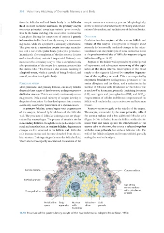
Corona radiata Corona radiata
Cortical granule
Cell of the
corona radiata
with cytoplasmic
Zona pellucida
process
Perivitelline Golgi Nucleus Mitochon- Lipid
space apparatus with drion granule
nucleolus
14.10 Structure of the postovulatory oocyte of the ewe (schematic; adapted from Rüsse, 1983).
Vet Histology.indb 308 16/07/2019 15:05