
Page 22 - Rapid Review of ECG Interpretation in Small Animal Practice, 2nd Edition
P. 22
Principles of Electrocardiography
VetBooks.ir Electrical properties of the heart
Two types of action potentials are observed in the heart: The fast response action potential occurs in the
normal atrial and ventricular myocardium and in the Purkinje fibers (Fig. 1.8A), while the slow response
action potential (Fig. 1.8B) is found in the SA node, the pacemaker region of the heart, and in the AV node,
the specialized tissue that conducts the cardiac impulse from atria to the ventricles (see Fig. 1.7, p. 5).
The various phases of the action potential (Fig. 1.8) are associated with changes in the permeability of the
+
2+
+
cell membrane, mainly to positively charged sodium (Na ), potassium (K ), and calcium (Ca ) ions. This is
accomplished by opening and closing of voltage-dependent ion channels in the cell membrane, selective
for individual ions. At rest (phase 4), the Purkinje fiber cells (Fig. 1.8A) maintain an electrical gradient
across the cell membranes (resting membrane potential) such that the inside is negative with respect to
+
the outside of the cells. This negative intracellular potential is maintained by Na channels, which extrude
Na ions from the cell. When an action potential from a neighboring cell arrives, it reduces the resting
+
potential to a threshold (i.e., makes it less negative) resulting in an abrupt increase in permeability of the
+
Na channels, allowing Na ions to rush into the cell and depolarize it. The rapid depolarization (upstroke of
+
fast response action potential), that ensues once the cell reaches a voltage threshold corresponds to
phase 0. The membrane potential is thus reversed or positive. Once a cell is depolarized, it cannot be
depolarized again, until the ionic fluxes that occur during depolarization are reversed, a process called
repolarization. The repolarization of the cardiac cells roughly corresponds to phases 1 through 3 of the
action potential. Phase 1 consists of a brief rapid repolarization, initiated at the end of the action potential
+
upstroke, as the Na channels inactivate and the K channels transiently allow an outward current. Phase
+
1 is interrupted when the cell reaches the “plateau phase” or phase 2, maintained by a slow Ca 2+ influx.
The transmembrane current of Ca initiates the mechanical contraction of the heart. During the plateau,
2+
2+
repolarization slowly progresses, until the Ca channels are turned off, and eventually a final rapid
+
repolarization (phase 3) ensues, via an outward K current. Because a second depolarization cannot happen
until repolarization occurs, the time from the end of phase 0 to late in phase 3 is called the refractory period.
Phase 4 is the resting membrane potential. This is the period that the cell remains in until it is stimulated
again by an external electrical stimulus (typically an action potential from an adjacent cell).
Cells from the SA node and the AV node (Fig. 1.8B) have a lower resting membrane potential which
becomes gradually more positive during diastole (phase 4) because of steady influx of calcium through
2+
slow Ca 2+ channels, eventually resulting in spontaneous depolarization. The slow influx of Ca produces
the slow upstroke velocity (slow response action potential) in the SA and AV nodal cells.
A B
+50 1
2
0
0
3 3
–50 Threshold
0 4 potential
4 4
–100 R
Resting
P T potential
Q
S
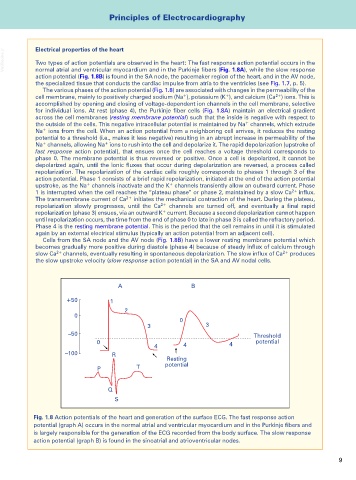
Fig. 1.8 Action potentials of the heart and generation of the surface ECG. The fast response action
potential (graph A) occurs in the normal atrial and ventricular myocardium and in the Purkinje fibers and
is largely responsible for the generation of the ECG recorded from the body surface. The slow response
action potential (graph B) is found in the sinoatrial and atrioventricular nodes.
9