
Page 39 - Rapid Review of ECG Interpretation in Small Animal Practice, 2nd Edition
P. 39
Approach to Evaluating Arrhythmias
ATRIAL STANDSTILL AV CONDUCTION ABNORMALITIES
(AV BLOCK)
Atrial standstill is a lack of electrocardiographic
VetBooks.ir evidence of atrial depolarization, resulting in the AV block consists of incomplete, intermittent, or
complete failure of conduction between the atria
absence of P waves on the ECG. Causes include
hyperkalemia, digitalis toxicity, and primary atrial and ventricles via the AV node. Three types of AV
myocardial disease. block exist:
• First-degree AV block (Fig. 3.10) is defined as
ECG criteria (Fig. 3.9): prolonged conduction through the AV node that
• No P waves are present. results in an increased PR interval of >0.13 s
• Slow junctional or ventricular escape rhythm is (dog) and >0.09 s (cat) and normal P wave and
often present. QRS complexes, in a 1:1 ratio.
• If associated with hyperkalemia, the T waves • Second-degree AV block (Fig. 3.11) is a
can be tall and the QRS morphology can be conduction disorder in which some atrial
wide and bizarre. impulses are not conducted to the ventricles.
There are normal P waves and QRS complexes,
but intermittently the P waves are not followed
by QRS complexes (blocked P). Second-degree
AV block occurs in two types:
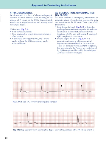
Fig. 3.9 Cat, lead aVL, 50 mm/s showing atrial standstill.
Fig. 3.10 Dog, lead II, 50 mm/s showing first-degree atrioventricular block, with a PR interval of 0.16 s.
26