
Page 432 - The Veterinary Care of the Horse
P. 432
VetBooks.ir
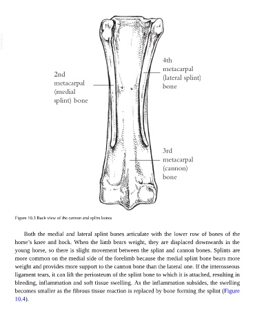
Figure 10.3 Back view of the cannon and splint bones
Both the medial and lateral splint bones articulate with the lower row of bones of the
horse’s knee and hock. When the limb bears weight, they are displaced downwards in the
young horse, so there is slight movement between the splint and cannon bones. Splints are
more common on the medial side of the forelimb because the medial splint bone bears more
weight and provides more support to the cannon bone than the lateral one. If the interosseous
ligament tears, it can lift the periosteum of the splint bone to which it is attached, resulting in
bleeding, inflammation and soft tissue swelling. As the inflammation subsides, the swelling
becomes smaller as the fibrous tissue reaction is replaced by bone forming the splint (Figure
10.4).