
Page 9 - NYSAGD GP Fall 2018
P. 9
Denture Related Pathology
By Gwen Cohen Brown, DDS, FAAOMP
Denture Associated Pathologies
Removable partial and full dentures are intended to restore esthet- Denture stomatitis may be present in up to 70% of full denture
ics and function to a patient with complete or partial edentulism. wearing patients with a slight female predilection. It is the most
However great their benefits, patients with removable dentures common form of oral candidiasis. Treatment includes proper
2
often develop specific denture associated pathologies. The most cleaning of dentures especially after meals and before bed, remov-
common pathologies are denture stomatitis, sometimes referred al of dentures at night, and treatment with antifungal therapy and/
to as denture sore mouth or papillary hyperplasia of the palate, or a disinfecting solution. If not treated, denture stomatitis can
epulis fissuratum, denture trauma (denture irritation hyperplasia/ lead to progressive bone atrophy, poorly fitting dentures as well as
ulceration), and angular cheilitis. Most of these conditions may be traumatic ulcers, epulis fissuratum and localized discomfort under
reduced or treated easily, although, depending upon the severity, the denture base. Treatment with Chlorhexidine, Amphotericin B,
4
new dentures may need to be fabricated following appropriate me- Listerine and Nystatin mouth rinse have all proven to be effective
dicinal or surgical treatment. methods of treatment, and meta-analysis has shown that disinfec-
tion, antiseptic and antifungals can all prove to be effective treat-
Denture Stomatitis ment when used appropriately. 4
The true etiology of denture stomatitis (denture sore mouth) is
multifactorial, however most researchers believe that candida albi- Traumatic Ulcers (Denture
cans plays a significant part in its development. Other factors may Associated Ulcers/Denture
include poor fitting dentures, certain bacterial species including Sore Spots)
staphylococcus species, streptococcus species, and neisseria spe- Traumatic ulcers (Figure 10) may
cies as well as local factors including wearing dentures through the develop with the continued use of
night and a carbohydrate rich diet. 1 unstable or ill-fitting dentures or
following the insertion of new par-
Denture stomatitis clinically presents as inflamed, red swollen mu- tial or full dentures, or may emerge
cosa directly subjacent to the denture base. It is considered to be over time secondary to bony atrophy
a type of candidiasis, chronic erythematous or chronic atrophic in under the denture base. Percentages
5
appearance. C albicans is a normal commensal organism of the range from 25% in patients wearing
mouth and generally does not cause problems in healthy people, complete dentures to 92% in patients Figure 10. Denture ulcer.
however, dentures often alter the normal oral microbiota. wearing a complete denture who
were also seeking a new denture
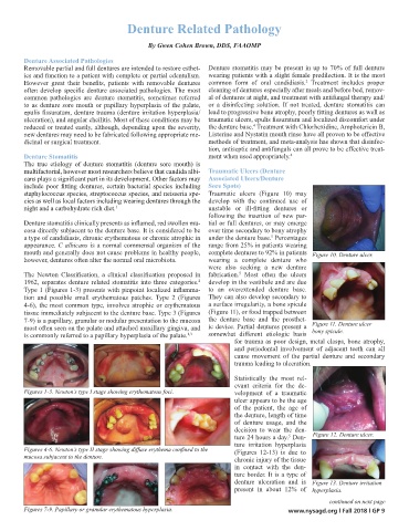
The Newton Classification, a clinical classification proposed in fabrication. Most often the ulcers
5
1962, separates denture related stomatitis into three categories. develop in the vestibule and are due
2
Type 1 (Figures 1-3) presents with pinpoint localized inflamma- to an overextended denture base.
tion and possible small erythematous patches. Type 2 (Figures They can also develop secondary to
4-6), the most common type, involves atrophic or erythematous a surface irregularity, a bone spicule
tissue immediately subjacent to the denture base. Type 3 (Figures (Figure 11), or food trapped between
7-9) is a papillary, granular or nodular presentation to the mucosa the denture base and the prosthet- Figure 11. Denture ulcer
most often seen on the palate and attached maxillary gingiva, and ic device. Partial dentures present a bony spicule.
is commonly referred to a papillary hyperplasia of the palate. 1,3 somewhat different etiologic basis
for trauma as poor design, metal clasps, bone atrophy,
and periodontal involvement of adjacent teeth can all
cause movement of the partial denture and secondary
trauma leading to ulceration.
Statistically the most rel-
evant criteria for the de-
Figures 1-3. Newton’s type I stage showing erythematous foci. velopment of a traumatic
ulcer appears to be the age
of the patient, the age of
the denture, length of time
of denture usage, and the
decision to wear the den-
ture 24 hours a day. Den- Figure 12. Denture ulcer.
5
ture irritation hyperplasia
Figures 4-6. Newton’s type II stage showing diffuse erythema confined to the (Figures 12-13) is due to
mucosa subjacent to the denture. chronic injury of the tissue
in contact with the den-
ture border. It is a type of
denture ulceration and is Figure 13. Denture irritation
present in about 12% of hyperplasia.
continued on next page
Figures 7-9. Papillary or granular erythematous hyperplasia. www.nysagd.org l Fall 2018 l GP 9