
Page 10 - NYSAGD GP Fall 2018
P. 10
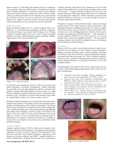
denture wearers.  Cheek biting and traumatic ulcers are commonly  cracking, fissuring, and redness at the commissures at one or both
                     6
        seen in patients with removable dentures. Treatment for traumatic  corners of the mouth and is a result of saliva pooling on the corners
        ulcers includes adjustment of the denture base for those patients  of the mouth. 13,14  Angular cheilitis can appear to be wet or dry, and
        who do not require relining or replacement of the dentures. Appli-  cracked with areas of bleeding. Most people find it to be both pain-
        cation of a mild topical steroid to the traumatic ulcer can help heal  ful and physically unattractive. While it had been associated with
        the ulceration and may be used in conjunction with altering the  nutritional defiency in the past it is currently thought to be due to
        denture base. Surgical excision of tissue associated with denture  infection with candida albicans. 15
        irritation hyperplasia or bony spicules may also be indicated. 7
                                                               Treatment of angular cheilitis should include a topical antifungal
        Epulis Fissuratum                                      possibly mixed with a mild steroid for better penetration of the
        Epulis  fissuratum  (Figures  14-17),  ‘denture-induced  fibrous  hy-  antifungal. Intraoral candidiasis should also be treated as angular
        perplasia’, is a soft tumor-like hyperplasia of fibrous connective  cheilitis and is often seen secondarily to intraoral candidiasis. Top-
        tissue that develops in association with the flange of an ill-fitting  ical polyene or azole antifungal agents are effective in most cases.
        complete or partial denture and is the result of bony resorption  Nystatin oral suspension (100,000 units/mL – 1 mL topically), or
        (atrophy) over time. Epulis fissuratum appears clinically as flabby  nystatin pastilles (100,000 IU) four times daily for 7 to 14 days
        ridges under a full or partial denture base.           should resolve most local candidal infections. 15,16  If the patient is
                                                               wearing a denture, they should be reminded to remove the denture
                                                               while treating the candida infection.
                                                               Conclusion
                                                               Patients with full or partial removable prosthetics should be mon-
                                                               itored for the development of these denture related pathologies.
                                                               Denture stomatitis, traumatic ulcers, epulis fissuratum, and angu-
                                                               lar cheilitis are the most commonly associated pathologic diseases
                                                               associated with long-term denture use. These conditions should be
                                                               treated as early as possible with appropriate measures, medicinal,
                                                               surgical denture alteration or if necessary fabrication of a new full
                                                               or partial denture.

                                                               All photographs were taken by Dr. Gwen Cohen Brown and are
                                                               courtesy of the Dental Hygiene Department of New York City Col-
                                                               lege of Technology.

                                                                   1.  Arendorf, T. M., and D. M. Walker. “Denture stomatitis: a re-
                                                                       view.” Journal of oral rehabilitation 14.3 (1987): 217-227.
                                                                   2.  Tyldesley, Anne Field, Lesley Longman in collaboration with
        Figures 14-17. Epulis fissuratum.                              William R. (2003). Tyldesley’s Oral medicine (5th ed.). Oxford:
                                                                       Oxford University Press. pp. 35–40. ISBN 0192631470.
        For the most part, patients are unaware of the excess tissues and   3.  Scully, Crispian (2008). Oral and maxillofacial medicine : the
        epulis  fissuratum  is  generally  asymptomatic.   Epulis  fissuratum   basis of diagnosis and treatment (2nd ed.). Edinburgh: Chur-
                                             8
        are generally seen in an older population, which is consistent with   chill Livingstone. pp. 201–203. ISBN 9780443068188.
        patients wearing removable dentures. However, there is a slight   4.  Emami, Elham, et al. “Linking evidence to treatment for den-
        female predilection suggesting that there may be a hormonal com-  ture stomatitis: a meta-analysis of randomized controlled tri-
        ponent as well.  Treatment is excision of the movable tissue either   als.” Journal of dentistry 42.2 (2014): 99-106.
                    9
        by surgical blade excision or by electro-cautery or laser. 10
        Etiology of epulis fissuratum is associated with both trauma and ir-
        ritation of the partial or full denture with the development of bony
        atrophy and subsequent growth of fibrous hyperplasia. The fibrous
        tissue may by ulcerated or secondarily infected with candida albi-
        cans.  Epulis fissuratum may result in the patient having difficulty
            11
        masticating and/or speaking correctly. Ectopic movements of the
        denture base may result in a more rapid destruction of the bony
        ridge so excision of excess movable tissue is indicated prior to
        fabricating a new denture. Treating the ulceration or candidal in-
        fection prior to excising the excess tissue may yield better results
        and faster healing.

        Angular Cheilitis
        Angular cheilitis (Figures 18-20) is often seen in patients wear-
        ing removable full or partial dentures in conjunction with intraoral
        candidiasis. It may be a result of reduced vertical dimension. It has
        also been associated with Vitamin B defiency, staphylococci and
        streptococci bacteria, diabetes and AIDS.  Angular cheilitis is a
                                          12
        chronic inflammatory condition that presents as an erythematous
        www.nysagd.org l Fall 2018 l GP 10                                 Figure 18-20. Angular cheilitis.