
Page 142 - Microsurgery
P. 142
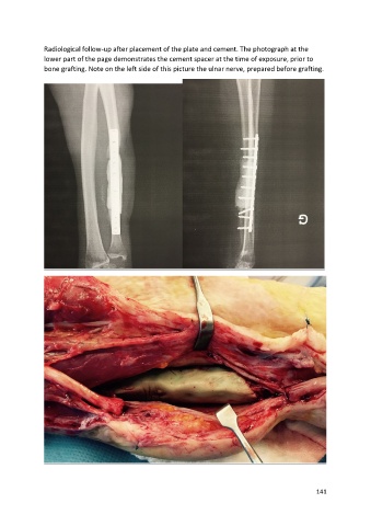
Radiological follow-up after placement of the plate and cement. The photograph at the
               lower part of the page demonstrates the cement spacer at the time of exposure, prior to
               bone grafting. Note on the left side of this picture the ulnar nerve, prepared before grafting.


                                                                                                       141