
Page 102 - Libro 2
P. 102
82
PART 2 — CEREBROVASCULAR
this, sonographers may not consider this a problem of concern. Still, a duplex evaluation is often the first diagnostic step and the sonographer must know of these abnormalities in order to perform an adequate assessment when evaluating a swollen neck.
Pitfalls
In the early postoperative period within the first few days after surgery, ultrasound visualization should be expected to be complicated by air entrapped in the matrix of a synthetic patch or introduced in the hemostatic agents applied prior to closure or by wound hematomas. Because entrapped air often obliterates the ultrasound image directly above the CEA site, the sonographer is often required to visual- ize the carotid bifurcation from the most posterior approach possible.
By the time of a first postoperative follow-up, the image is no longer compromised by entrapped air. Wound hematomas should have been substantially reduced. Nonreduction and tenderness should be considered particularly problematic in the presence of a swollen neck.
DIAGNOSIS
Intravascular problems associated with the CEA may include both stenotic or nonstenotic pathology. One type of nonstenotic issue associated with the CEA includes an oversized or irregular patch that gives the vessel an aneurysmal appearance. Diameters are important measurements in these cases. It should be remembered that a patch identified as synthetic is more thrombogenic than an autogenous patch. Slower flows in an aneurysmal patch are more likely
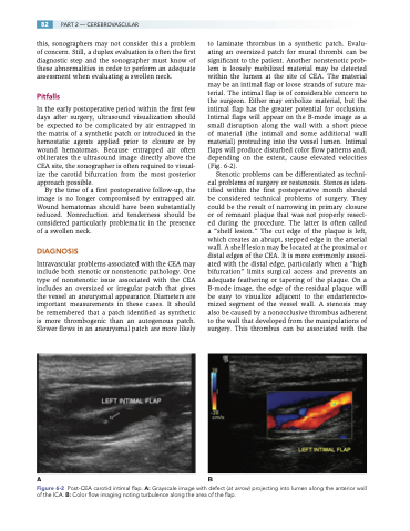
to laminate thrombus in a synthetic patch. Evalu- ating an oversized patch for mural thrombi can be significant to the patient. Another nonstenotic prob- lem is loosely mobilized material may be detected within the lumen at the site of CEA. The material may be an intimal flap or loose strands of suture ma- terial. The intimal flap is of considerable concern to the surgeon. Either may embolize material, but the intimal flap has the greater potential for occlusion. Intimal flaps will appear on the B-mode image as a small disruption along the wall with a short piece of material (the intimal and some additional wall material) protruding into the vessel lumen. Intimal flaps will produce disturbed color flow patterns and, depending on the extent, cause elevated velocities (Fig. 6-2).
Stenotic problems can be differentiated as techni- cal problems of surgery or restenosis. Stenoses iden- tified within the first postoperative month should be considered technical problems of surgery. They could be the result of narrowing in primary closure or of remnant plaque that was not properly resect- ed during the procedure. The latter is often called a “shelf lesion.” The cut edge of the plaque is left, which creates an abrupt, stepped edge in the arterial wall. A shelf lesion may be located at the proximal or distal edges of the CEA. It is more commonly associ- ated with the distal edge, particularly when a “high bifurcation” limits surgical access and prevents an adequate feathering or tapering of the plaque. On a B-mode image, the edge of the residual plaque will be easy to visualize adjacent to the endarterecto- mized segment of the vessel wall. A stenosis may also be caused by a nonocclusive thrombus adherent to the wall that developed from the manipulations of surgery. This thrombus can be associated with the
AB
Figure 6-2 Post-CEA carotid intimal flap. A: Grayscale image with defect (at arrow) projecting into lumen along the anterior wall of the ICA. B: Color flow imaging noting turbulence along the area of the flap.
