
Page 103 - Libro 2
P. 103
CEA site and patch.4 Lastly, although the post-CEA exam is targeted at the operative site, there is still the possibility of missed lesions in the proximal CCA or the innominate artery (on the right) that had been overlooked in the presurgical workup.
Restenosis
Although a stenosis seen 1 month postsurgery is typi- cally associated with surgery in the form of retained plaque or thrombus, narrowing in the first 24 months after a CEA is considered a result of neointimal hy- perplasia. The hyperplastic lesion is considered rela- tively benign with the low thromboembolic potential of a fibrotic plaque. After 2 years, a stenosis is con- sidered atherosclerotic and potentially more problem- atic, with risks approaching that of the asymptomatic lesion. Restenosis rates vary widely in the literature, but Rosenborough and Perler estimate the incidence of restenosis as 6% to 14% after a CEA.5
When evaluating the patient in follow-up, some laboratories may adjust velocity criteria post-CEA. Most will use the preprocedural velocity criteria established by the lab to identify the presence of a stenosis. Sonographers may question the need for postoperative surveillance if the restenosis rate is relatively low and the lesion is relatively benign. In some respects, it matches the surveillance of the asymptomatic lesion. Unlike the primary asymptom- atic lesion, however, the hyperplastic response to surgery has the potential to become virulent with a rapid progression to occlusion. The primary aims of the surgeon are to identify the restenosis early and, when the disease is found to progress in follow-up, to intervene before it reaches occlusion. Follow-up is also important for the contralateral bifurcation. Atherogenesis has a degree of symmetry. Patients treated for disease in one carotid bifurcation are at risk of aggressive disease in the contralateral bifur- cation. Postoperative surveillance should always be performed bilaterally (Pathology Box 6-1).
CAROTID ARTERY STENTING
In 2005, carotid artery stenting (CAS) accounted for only 9.6% of patients treated for extracranial cere- brovascular disease.6 Held back by concerns of high complication rates and low reimbursements, CAS has not benefitted from wide use. Complication rates for CAS continue to decrease with the increased use of cerebral protective devices and retrograde flow flushing of the ICA during stenting.
The rapid evolution of CAS since 2005 can be dem- onstrated in three publications. In 2008, the Society for Vascular Surgery (SVS) published guidelines for
the management of carotid stenosis.7 It was recom- mended that CEA be used for the moderate-to-severe symptomatic carotid stenoses and asymptomatic lesions. CAS was only recommended for symptom- atic patients with high perioperative risk and not for asymptomatic stenoses.
In 2010, the Carotid Revascularization Endarter- ectomy vs. Stenting Trial (CREST) reported its long awaited results.8 Although preliminary results dem- onstrated that CAS patients suffered more strokes than CEA patients in the 30 days postprocedure, this finding was balanced by the number of heart attacks in the CEA group. Within the immediate postopera- tive period, there was no statistical difference be- tween CAS and CEA in heart attacks and strokes. This equivalence was found to continue over the
6 — Ultrasound Following Surgery and Intervention 83
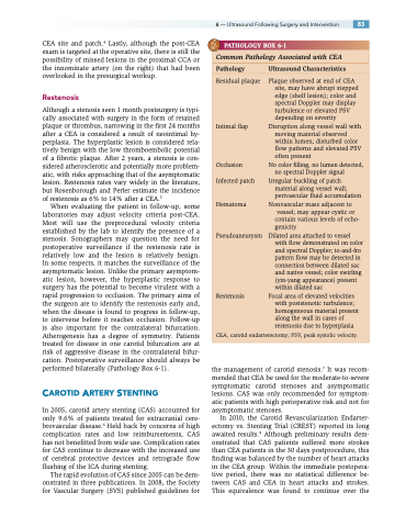
PATHOLOGY BOX 6-1
Common Pathology Associated with CEA
Pathology UltrasoundCharacteristics
Residual plaque
Intimal flap
Occlusion Infected patch
Hematoma
Pseudoaneurysm
Restenosis
Plaque observed at end of CEA site, may have abrupt stepped edge (shelf lesion); color and spectral Doppler may display turbulence or elevated PSV depending on severity
Disruption along vessel wall with moving material observed within lumen; disturbed color flow patterns and elevated PSV often present
No color filling, no lumen detected, no spectral Doppler signal
Irregular buckling of patch material along vessel wall; perivascular fluid accumulation
Nonvascular mass adjacent to vessel; may appear cystic or
contain various levels of echo-
genicity
Dilated area attached to vessel
with flow demonstrated on color and spectral Doppler; to-and-fro pattern flow may be detected in connection between dilated sac and native vessel; color swirling (yin-yang appearance) present within dilated sac
Focal area of elevated velocities with poststenotic turbulence; homogeneous material present along the wall in cases of restenosis due to hyperplasia
CEA, carotid endarterectomy; PSV, peak systolic velocity.
