
Page 105 - Libro 2
P. 105
6 — Ultrasound Following Surgery and Intervention
85
AB
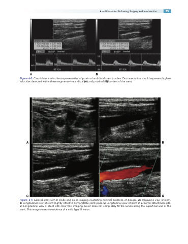
Figure 6-3 Carotid stent velocities representative of proximal and distal stent borders. Documentation should represent highest velocities detected within these segments—near distal (A) and proximal (B) borders of the stent.
AB
CD
Figure 6-4 Carotid stent with B-mode and color imaging illustrating minimal evidence of disease. A: Transverse view of stent. B: Longitudinal view of stent slightly offset to demonstrate stent walls. C: Longitudinal view of stent at proximal attachment site. D: Longitudinal view of stent with color flow imaging. Color does not completely fill the lumen along the superficial wall of the stent. This image serves as evidence of a mild Type III lesion.
