
Page 106 - Libro 2
P. 106
86 PART 2 — CEREBROVASCULAR Pitfalls
Dense calcification that produced shadows in the preprocedural scan will be present in the follow-up evaluation and will compromise the B-mode image and Doppler interrogation of the stented vessel. Mul- tiple views should be used to avoid areas of acous- tic shadowing. In some vessels, this is not possible; therefore, signals distal to the calcific areas will be important in determining the presence of disease. Turbulence distal to these areas likely indicates that a stenosis is present.
Dense circumferential calcification is problematic during the stent placement as well. It restricts bal- loon expansion of the lesion during stenting, which in turn increases procedural manipulation and adds the risk of an inadequate expansion. Both are rea- sons for sonographers to pay particular attention to velocity changes with the area of calcification. Added manipulation may increase the hyperplastic response, and inadequate expansion may lead to re- sidual stenosis.
DIAGNOSIS
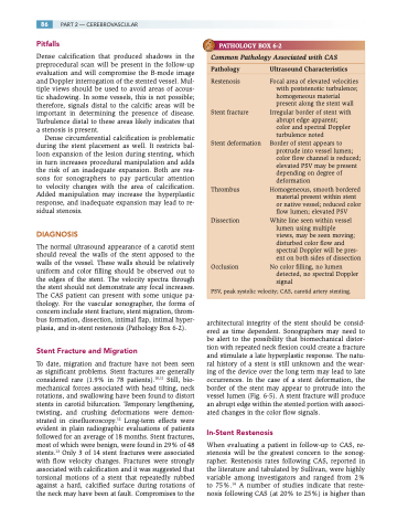
The normal ultrasound appearance of a carotid stent should reveal the walls of the stent apposed to the walls of the vessel. These walls should be relatively uniform and color filling should be observed out to the edges of the stent. The velocity spectra through the stent should not demonstrate any focal increases. The CAS patient can present with some unique pa- thology. For the vascular sonographer, the forms of concern include stent fracture, stent migration, throm- bus formation, dissection, intimal flap, intimal hyper- plasia, and in-stent restenosis (Pathology Box 6-2).
Stent Fracture and Migration
To date, migration and fracture have not been seen as significant problems. Stent fractures are generally considered rare (1.9% in 78 patients).10,11 Still, bio- mechanical forces associated with head tilting, neck rotations, and swallowing have been found to distort stents in carotid bifurcation. Temporary lengthening, twisting, and crushing deformations were demon- strated in cinefluoroscopy.12 Long-term effects were evident in plain radiographic evaluations of patients followed for an average of 18 months. Stent fractures, most of which were benign, were found in 29% of 48 stents.13 Only 3 of 14 stent fractures were associated with flow velocity changes. Fractures were strongly associated with calcification and it was suggested that torsional motions of a stent that repeatedly rubbed against a hard, calcified surface during rotations of the neck may have been at fault. Compromises to the
architectural integrity of the stent should be consid- ered as time dependent. Sonographers may need to be alert to the possibility that biomechanical distor- tion with repeated neck flexion could create a fracture and stimulate a late hyperplastic response. The natu- ral history of a stent is still unknown and the wear- ing of the device over the long term may lead to late occurrences. In the case of a stent deformation, the border of the stent may appear to protrude into the vessel lumen (Fig. 6-5). A stent fracture will produce an abrupt edge within the stented portion with associ- ated changes in the color flow signals.
In-Stent Restenosis
When evaluating a patient in follow-up to CAS, re- stenosis will be the greatest concern to the sonog- rapher. Restenosis rates following CAS, reported in the literature and tabulated by Sullivan, were highly variable among investigators and ranged from 2% to 75%.14 A number of studies indicate that reste- nosis following CAS (at 20% to 25%) is higher than
PATHOLOGY BOX 6-2
Common Pathology Associated with CAS
Pathology UltrasoundCharacteristics
Restenosis
Stent fracture
Stent deformation
Thrombus
Dissection
Occlusion
Focal area of elevated velocities with poststenotic turbulence; homogeneous material present along the stent wall
Irregular border of stent with abrupt edge apparent; color and spectral Doppler turbulence noted
Border of stent appears to protrude into vessel lumen; color flow channel is reduced; elevated PSV may be present depending on degree of deformation
Homogeneous, smooth bordered material present within stent or native vessel; reduced color flow lumen; elevated PSV
White line seen within vessel lumen using multiple
views, may be seen moving; disturbed color flow and spectral Doppler will be pres- ent on both sides of dissection
No color filling, no lumen detected, no spectral Doppler signal
PSV, peak systolic velocity; CAS, carotid artery stenting.
