
Page 108 - Libro 2
P. 108
88
PART 2 — CEREBROVASCULAR
TABLE 6-2
Post-CAS Duplex Ultrasound Criteria
STENOSIS THRESHOLD
ABURAHMA17
SETACCI18
CHI23
CHAHWAN22
LAL20
ZHOU19
ARMSTRONG24
PSV
EDV
PSV
EDV
I/C
PSV
I/C
PSV
EDV
PSV
I/C
PSV
EDV
I/C
PSV
EDV
20%
137
20
150
2.15
30%
154
42
105
50%
224
88
175
240
2.45
195
62
220
2.7
70%
300
140
3.8
450
4.3
300
90
4.0
75%
300
125
80%
325
119
300
96
340
4.15
PSV, peak systolic velocity; EDV, end-diastolic velocity; I/C, ICA/CCA PSV ratio.
In Table 6-2, the primary discriminator for a sig- nificant stenosis is the peak systolic velocity (PSV). End-diastolic velocity (EDV) did not appear as dis- criminating,20,23 with the exception of one study.24 The ICA/CCA PSV ratio, although reported, did not appear to substantially add to the determination of restenosis.
The PSV threshold for the 50% stenosis varied from 175 to 240 cm/s among criteria, suggesting it may not be a good discriminator for moderate steno- sis. It was noted in the literature and verified by this range of velocities that velocity was not uniformly el- evated among all patients. This suggests that PSV may not provide a reliable estimate for moderate stenosis.
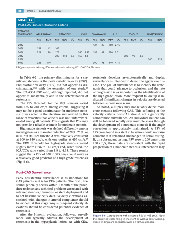
High-grade stenosis was defined differently among investigators as a diameter reduction of 70%, 75%, or 80% but its PSV threshold was relatively consistent at 300 to 340 cm/s, with one outlier at 450 cm/s.24 The EDV threshold for high-grade stenosis varied slightly more at 96 to 140 cm/s and, when used, the ICA/CCA ratio varied from 3.8 to 4.15. These results suggest that a PSV of 300 to 325 cm/s could serve as a relatively good predictor of a high-grade restenosis (Fig. 6-6).
Post-CAS Surveillance
Early poststenting surveillance is as important for CAS patients as it is for CEA patients. The first ultra- sound generally occurs within 1 month of the proce- dure to detect any technical problems associated with retained stenosis, thrombus, or stent deployment and to set baseline velocity data. Velocity elevations as- sociated with changes in arterial compliance should be evident at this stage. Any subsequent velocity el- evations should be considered potential evidence of restenosis.
After the 1-month evaluation, follow-up surveil- lance will typically address the development of restenosis in the hyperplastic lesion. As with CEA,
restenosis develops asymptomatically and duplex surveillance is intended to detect the aggressive dis- ease. The goal of surveillance is to identify the reste- nosis that could advance to occlusion, and the rate of progression is as important as the identification of the high-grade lesion. More frequent follow-up is in- dicated if significant changes in velocity are detected between surveillance scans.
As noted, a duplex may not reliably detect mod- erate stenosis following CAS. This softening of the velocity criteria post-CAS should not significantly compromise surveillance. An individual patient can still be followed serially over multiple scans through the development of a moderate stenosis if the angle correction is appropriately maintained. A PSV of 175 cm/s found in a stent at baseline should not raise concerns if it remained unchanged in serial testing. If, on subsequent testing, PSV rose to 200 cm/s then 250 cm/s, these data are consistent with the rapid progression of a moderate stenosis. Intervention may
Figure 6-6 Carotid stent with elevated PSV at 405 cm/s. Note the narrowed color filling in the stent as well as color aliasing. These findings are consistent with a 70% restenosis.
