
Page 107 - Libro 2
P. 107
following CEA. As with CEA, restenosis is consid- ered to be a low-risk neointimal hyperplastic lesion with an occurrence within the first 2 years of stent- ing. A difference in the hyperplastic response may, however, lie in the type of insult. Hyperplasia with CEA is considered an early occurrence in response to a single insult. De Borst et al. suggests that the hy- perplasia following CAS may be an ongoing response to the presence of an implanted foreign object.15 Giv- en these findings, follow-up surveillance may appear more significant for CAS than for CEA.
Lal et al. characterized the in-stent restenosis (ISR) in five sonographic distribution patterns (Table 6-1).16 All ISRs are described as intimal hyperplasia with its typical homogeneously hypoechoic appear- ance. The most common form of ISR (Type I) de- veloped at the stent border and comprised 40% of the restenosis that were identified by B-mode and color/power Doppler. The most severe was occlusion (Type V), which was rare at 1.2%. The second most severe form of ISR, Type IV, was diffuse proliferative narrowing of the stent lumen. Type IV lesions were found in 20% of ISRs and was most predictive of the
TABLE 6-1
Classification of In-stent Restenosis (ISR)16
need for reintervention. On an interesting note, Lal et al. found that Type IV ISR tend to be associated with diabetes and concurred with other investiga- tors that diabetes is a predictor of aggressive intimal hyperplasia.16
Changes in Velocity Criteria
Although increased ISR rates were reported with stents over CEA, other concurrent studies have doc- umented that flow velocity elevations occur in post- CAS patients without the presence of stenosis. It was considered that the stent may impose a rigid matrix that reduces arterial compliance and decreases the elastic expansion of the stented segment. The result may be an elevation in velocities within the stent that could be misinterpreted as a restenosis.
Flow velocity elevations have lead investigators to propose a number of new velocity criteria to use when evaluating the post-CAS patient.17–24 Table 6-2 compares the proposed criteria. All elevate the ve- locity thresholds for the moderate and high-grade stenosis.
6 — Ultrasound Following Surgery and Intervention 87
AB
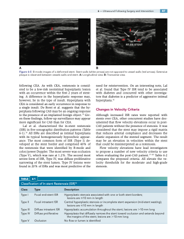
Figure 6-5 B-mode images of a deformed stent. Stent walls (white arrows) are not apposed to vessel walls (red arrows). Extensive plaque is observed between vessels walls and stent. A: Longitudinal view. B: Transverse view.
Class Type
Description
Type I
Type II
Type III Type IV
Type V
Focal end-stent ISR Focal intrastent ISR
Diffuse intrastent ISR Diffuse proliferative
Occlusion
Hyperplastic stenosis associated with one or both stent borders; lesions are 10 mm in length
Central hyperplastic stenosis or incomplete stent expansion (mid-stent wasting); lesions are 10 mm in length
Hyperplastic accumulation throughout the stent; lesions are 10 mm long Hyperplasia that diffusely narrows the stent toward occlusion and extends beyond
the margins of the stent; lesions are 10 mm long No flow or lumen is identified
