
Page 119 - Libro 2
P. 119
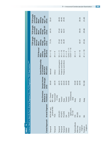
7 — Intracranial Cerebrovascular Examination 99
TABLE 7-2
Criteria Used to Identify Each Arterial Segment for a Full Diagnostic TCD Examination18,19
Cranial Approach
Arterial Segment
Age 20–39 (cm/s)
Age 40–59 (cm/s)
Age 60 (cm/s)
Temporal Temporal
MCA (M1 and proximal M2)
M1 – Toward M2 – Away
30–60 60–70
Identical
55 /12
71–76
69–76
55–61
Temporal Temporal Temporal Orbital Orbital
ACA (A1)
Away Toward Away Toward
60–75 60–75 60–65 35–55 65–80
Anterior and superior Posterior and inferior Posterior and inferior
50 /11
57–62 51–55 45–49
57–64 48–51 46–51
48–54 40–45 39–45
Submandibular Atlas loop Foramen
ICA
VA (V3) VA
Away
35–80 40–50 60–90
30 /9
38/10 42–47
magnum
TICA
Towardand/ or away
Inferior
39/9
PCA (P1)
39 /10
PCA (P2)
40 /10
OA
21/5
Carotid siphon (C4, C3, C2)
Toward, bidirectional, away
C2 41 /11 C4 47 /14
Foramen BA Away 70–120 magnum
41/10 47–53
Flow Direction Relative to Transducer
Sample Volume Depth Ranges (mm)
Spatial Relationship to Landmark MCA/ACA Bifurcation
TCD Normal Mean Velocity Range and SD (cm/s)
TCDI (angle corrected) Normal Mean Velocity Range
TCDI (angle corrected) Normal Mean Velocity Range
TCDI (angle corrected) Normal Mean Velocity Range
Away
38–43 39–48
30–36 31–40
