
Page 120 - Libro 2
P. 120
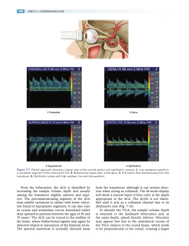
 100
 PART 2 — CEREBROVASCULAR
   4
2
3
1
    1 Parasellar
2 Genu
  3 Supraclinoid
4 Ophthalmic
Figure 7-7 Orbital approach showing a lateral view of the carotid siphon and ophthalmic arteries. 1: Low resistance waveform in parasellar segment of the intracranial ICA. 2: Bidirectional signal seen at the genu. 3: ICA siphon flow directed away from the transducer. 4: Ophthalmic artery with high resistant, low velocity waveform.
From the bifurcation, the ACA is identified by increasing the sample volume depth and usually aiming the transducer slightly anterior and supe- rior. The precommunicating segment of the ACA does exhibit variations in caliber with lower veloci- ties found in hypoplastic segments. It can also vary its course and sometimes curves downward rather than upward in patients between the ages of 50 and 70 years.2 The ACA can be traced to the midline of the brain, where bidirectional signals may again be detected related to insonation of the bilateral ACAs. The spectral waveform is normally directed away
from the transducer, although it can reverse direc- tion when acting as collateral. The M-mode display will show a narrow band of blue color at the depth appropriate to the ACA. The ACOA is not identi- fied until it acts as a collateral channel due to its diminutive size (Fig. 7-10).
To identify the TICA, the sample volume depth is returned to the landmark bifurcation and, at the same depth, aimed directly inferior. Velocities may appear low due to the anatomical course of the TICA relative to the sound beam, which tends to be perpendicular to the vessel, creating a larger
