
Page 122 - Libro 2
P. 122
102
PART 2 — CEREBROVASCULAR
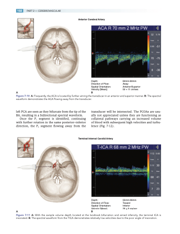
Anterior Cerebral Artery
Depth:
Direction of Flow: Spatial Orientation: Velocity [Mean]:
AB
60mm-80mm Away Anterior/Superior 50 + 11 cm/sec
Figure 7-10 A: Frequently, the ACA is located by further aiming the transducer in an anterior and superior manner. B: The spectral waveform demonstrates the ACA flowing away from the transducer.
left PCA are seen as they bifurcate from the tip of the BA, resulting in a bidirectional spectral waveform.
Once the P1 segment is identified, continuing with further rotation in the same posterior–inferior direction, the P2 segment flowing away from the
transducer will be intersected. The PCOAs are usu- ally not appreciated unless they are functioning as collateral pathways carrying an increased volume of blood with subsequent high velocities and turbu- lence (Fig. 7-12).
Terminal Internal Carotid Artery
Depth:
Direction of Flow: Spatial Orientation: Velocity [Mean]:
AB
55mm-65mm Toward Inferior
39 ± 9 cm/sec
Figure 7-11 A: With the sample volume depth located at the landmark bifurcation and aimed inferiorly, the terminal ICA is insonated. B: The spectral waveform from the TICA demonstrates relatively low velocities due to the poor angle of insonation.
