
Page 123 - Libro 2
P. 123
7 — Intracranial Cerebrovascular Examination
103
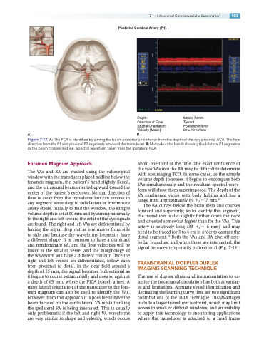
Posterior Cerebral Artery (P1)
AB
Figure 7-12 A: The PCA is identified by aiming the beam posterior and inferior from the depth of the very proximal ACA. The flow
direction from the P1 and proximal P2 segments is toward the transducer. B: M-mode color bands showing the bilateral P1 segments as the beam crosses midline. Spectral waveform taken from the ipsilateral PCA.
Foramen Magnum Approach
The VAs and BA are studied using the suboccipital window with the transducer placed midline below the foramen magnum, the patient’s head slightly flexed, and the ultrasound beam oriented upward toward the center of the patient’s eyebrows. Normal direction of flow is away from the transducer but can reverse in any segment secondary to subclavian or innominate artery steals. Initially to find the window, the sample volume depth is set at 60 mm and by aiming minimally to the right and left toward the orbit of the eye signals are found. The right and left VAs are differentiated by having the signal drop out as one moves from side to side and because the waveforms frequently have a different shape. It is common to have a dominant and nondominant VA, and the flow velocities will be lower in the smaller vessel and the morphology of the waveform will have a different contour. Once the right and left vessels are differentiated, follow each from proximal to distal. In the near field around a depth of 55 mm, the signal becomes bidirectional as it begins to course extracranially and does so again at a depth of 65 mm, where the PICA branch arises. A more lateral orientation of the transducer to the fora- men magnum can also be used to identify the VAs. However, from this approach it is possible to have the beam focused on the contralateral VA while thinking the ipsilateral VA is being insonated. This is usually only problematic if the left and right VA waveforms are very similar in shape and velocity, which occurs
about one-third of the time. The exact confluence of the two VAs into the BA may be difficult to determine with nonimaging TCD. In some cases, as the sample volume depth increases it begins to encompass both VAs simultaneously and the resultant spectral wave- form will show them superimposed. The depth of the VA confluence varies with body habitus and has a range from approximately 69 / 7 mm.20
The BA curves below the brain stem and courses forward and superiorly; so to identify this segment, the transducer is slid slightly further down the neck and oriented somewhat higher than for the VAs. This artery is relatively long (33 / 6 mm) and may need to be traced for 3 to 4 cm in order to capture the distal segment.19 Both the VAs and BA give off cere- bellar branches, and when these are intersected, the signal becomes temporarily bidirectional (Fig. 7-13).
TRANSCRANIAL DOPPLER DUPLEX IMAGING SCANNING TECHNIQUE
The use of duplex ultrasound instrumentation to ex- amine the intracranial circulation has both advantag- es and limitations. Accurate vessel identification and decreasing the learning curve time are two significant contributions of the TCDI technique. Disadvantages include a larger transducer footprint, which may limit access to small or difficult windows, and an inability to apply this technology to monitoring applications where the transducer is attached to a head frame
Depth:
Direction of Flow: Spatial Orientation: Velocity [Mean]:
60mm-70mm Toward Posterior/Inferior 39 ± 10 cm/sec
