
Page 124 - Libro 2
P. 124
104
PART 2 — CEREBROVASCULAR
Vertebral Artery
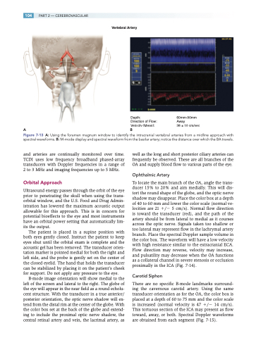
Depth:
Direction of Flow: Velocity [Mean]:
AB
60mm-90mm Away
38 ± 10 cm/sec
Figure 7-13 A: Using the foramen magnum window to identify the intracranial vertebral arteries from a midline approach with spectral waveforms. B: M-mode display and spectral waveform from the basilar artery; notice the distance over which the BA travels.
and arteries are continually monitored over time. TCDI uses low frequency broadband phased-array transducers with Doppler frequencies in a range of 2 to 3 MHz and imaging frequencies up to 5 MHz.
Orbital Approach
Ultrasound energy passes through the orbit of the eye prior to penetrating the skull when using the trans- orbital window, and the U.S. Food and Drug Admin- istration has lowered the maximum acoustic output allowable for this approach. This is in concern for potential bioeffects to the eye and most instruments have an orbital power setting that automatically lim- its the output.
The patient is placed in a supine position with both eyes gently closed. Instruct the patient to keep eyes shut until the orbital exam is complete and the acoustic gel has been removed. The transducer orien- tation marker is pointed medial for both the right and left side, and the probe is gently set on the center of the closed eyelid. The hand that holds the transducer can be stabilized by placing it on the patient’s cheek for support. Do not apply any pressure to the eye.
B-mode image orientation will show medial to the left of the screen and lateral to the right. The globe of the eye will appear in the near field as a round echolu- cent structure. With the transducer in a true anterior/ posterior orientation, the optic nerve shadow will ex- tend from the distal rim at the center of the globe. With the color box set at the back of the globe and extend- ing to include the proximal optic nerve shadow, the central retinal artery and vein, the lacrimal artery, as
well as the long and short posterior ciliary arteries can frequently be observed. These are all branches of the OA and supply blood flow to various parts of the eye.
Ophthalmic Artery
To locate the main branch of the OA, angle the trans- ducer 15% to 20% and aim medially. This will dis- tort the round shape of the globe, and the optic nerve shadow may disappear. Place the color box at a depth of 40 to 60 mm and lower the color scale (normal ve- locities are 21 / 5 cm/s). Normal flow direction is toward the transducer (red), and the path of the artery should be from lateral to medial as it courses across the optic nerve. Signals taken too shallow or too lateral may represent flow in the lachrymal artery branch. Place the spectral Doppler sample volume in the color box. The waveform will have a low velocity with high resistance similar to the extracranial ECA. Flow direction may reverse, velocity may increase, and pulsatility may decrease when the OA functions as a collateral channel in severe stenosis or occlusion proximally in the ICA (Fig. 7-14).
Carotid Siphon
There are no specific B-mode landmarks surround- ing the cavernous carotid artery. Using the same transducer orientation as for the OA, the color box is placed at a depth of 60 to 75 mm and the color scale is increased (normal velocity is 47 / 14 cm/s). This tortuous section of the ICA may present as flow toward, away, or both. Spectral Doppler waveforms are obtained from each segment (Fig. 7-15).
