
Page 133 - Libro 2
P. 133
7 — Intracranial Cerebrovascular Examination
113
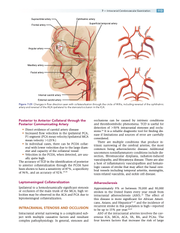
Supraorbital artery Frontal artery
Angular artery
Maxillary artery Facial artery
Internal carotid artery External carotid artery
Ophthalmic artery
Superficial temporal artery
Figure 7-25 Changes in flow direction seen with collateralization through the circle of Willis, including reversal of the ophthalmic artery and reversal of the ACA ipsilateral to the stenosis/occlusion in the ICA.
Posterior to Anterior Collateral through the Posterior Communicating Artery
• Direct evidence of carotid artery disease
• Increased flow velocities in the ipsilateral PCA, P1 segment (PCA mean velocity/ipsilateral MCA
mean velocity 125%)
• In individual cases, there can be PCOA collat-
eral with lower velocities due to the large diam-
eter and capacity of the collateral vessel
• Velocities in the PCOA, when detected, are usu-
ally quite high
The accuracy of TCD in the identification of posterior to anterior collateralization through the PCOA have been shown to have a sensitivity of 87%, a specificity of 96%, and an accuracy of 92%.23,24
Leptomeningeal Collateralization
Ipsilateral to a hemodynamically significant stenosis or occlusion of the main trunk of the MCA, high ve- locities may be observed in the ACA and PCA due to leptomeningeal collateralization.
INTRACRANIAL STENOSIS AND OCCLUSION
Intracranial arterial narrowing is a complicated sub- ject with multiple causative factors and resultant complex pathophysiology. In general, stenoses and
occlusions can be caused by intrinsic conditions and thromboembolic phenomena. TCD is useful for detection of 50% intracranial stenoses and occlu- sions.24 It is a reliable diagnostic tool for finding dis- ease if limitations and sources of error are carefully considered.
There are multiple conditions that produce in- trinsic narrowing of the cerebral arteries, the most common being atherosclerotic disease. Additional uncommon noninflammatory conditions include dis- section, fibromuscular dysplasia, radiation-induced vasculopathy, and Moyamoya disease. There are also a host of inflammatory vasculopathies and hemato- logic causes of stroke that may affect the basal cere- bral vessels including temporal arteritis, meningitis, toxin-related vasculitis, and sickle cell disease.
Atherosclerosis
Approximately 9% or between 70,000 and 90,000 strokes in the United States every year result from intracranial atherosclerosis (ASO).25 The effect of this disease is more significant for African Ameri- cans, Asians, and Hispanics25–27 and the incidence of recurrent stroke in this population is high—reported to be up to 15% per year.28,29
ASO of the intracranial arteries involves the cav- ernous ICA, MCA, ACA, VA, BA, and PCAs. The four known factors that increase the risk of large
