
Page 135 - Libro 2
P. 135
Embolic Stenosis and Occlusion
The most common cause of occlusion beyond the circle of Willis is embolism, accounting for 15% to 30% of strokes, most of which occur in the territory of the MCA.34 Several types of cardiac disease lead to cerebral embolism causing stenosis and occlu- sion including cardiac arrhythmias, ischemic heart disease, valvular disease, dilated cardiomyopathies, atrial septal abnormalities, and intracardiac tumors. Other sources of emboli include aortic arch athero- ma, extracranial carotid and VA plaque, and crossing of a venous thrombus into the arterial tree in pa- tients with patent foramen ovale.31
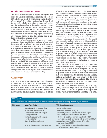
The use of cerebrovascular ultrasound in acute stroke requires a modified protocol that allows rapid insonation of the affected territory, supply arteries, and quick interpretation of the data. TCD can pro- vide significant information regarding a thrombus in acute intracranial arterial occlusion, often a dynamic process that can involve recanalization. A TCD flow grading system was developed by Demchuk to predict the success of intracranial clot lysis and short-term improvement after ischemic stroke. Thrombolysis in brain ischemia (TIBI) measures residual flow around the clot and, in general, a larger amount of residual flow predicts the success of the thrombolysis.35 For acute thrombosis, the TIBI scale is used to classify changes that can occur rapidly with recanalization and reocclusion in acute stroke (Table 7-4).
VASOSPASM
SAH, one of the most devastating types of stroke, accounts for 5% to 15% of all strokes and is fatal or disabling in about 60% of patients.36,37 These patients suffer the initial effect of an intracranial bleed, the risks and complications associated with surgical or interventional treatments, and the sequelae of a host
TABLE 7-4
of medical complications. One of the most signifi- cant causes of delayed ischemic neurological deficits (DINDS) is the development of cerebral vasospasm during the first 2-week period following the initial bleed. The pathophysiology, diagnosis, prevention, and treatment of vasospasm continue to be topics of intense investigation aimed at improving clinical outcomes for these patients.
Cerebral vasospasm is the transient and delayed narrowing of the basal cerebral arteries following SAH, and the exact cause remains the subject of in- tense study. It is mainly seen in the large skull base arteries and, less frequently, in the distal branches of these vessels, and it is responsible for the signifi- cant morbidity and mortality seen in this population. The onset of arterial contraction, as demonstrated by angiography, begins 3 to 4 days following the ini- tial bleed; peak narrowing develops at 6 to 8 days; and resolution occurs at 2 to 4 weeks post-SAH.38 The incidence of angiographic vasospasm after SAH is greater than 50%, with symptomatic vasospasm affecting one-third of all aneurysmal SAH patients.39 Neurological deficits caused by cerebral vasospasm may resolve or progress to infarction or death in spite of maximal therapy.
The medical management of cerebral vasospasm relies on hemodynamic therapy to improve cerebral blood flow. Calcium channel antagonists are widely used and have been shown to reduce poor outcomes. In patients with a clinically significant vasospasm that does not respond to maximal medical therapy, balloon angioplasty has been shown to be effec- tive, improving neurological deficits. The timing of angioplasty is significant and patients need to be treated before cerebral infarction occurs.
In 2004, the American Academy of Neurology (AAN) published a special article addressing the use of TCD and TCDI for the diagnosis of multiple intra- cranial vascular abnormalities according to a rating
TIBI Scale for TCD Detection of MCA Recanalization during and following Thrombolytic Therapy
7 — Intracranial Cerebrovascular Examination 115
TIBI Score
0 1 2
3 4 5
MCA Flow (DSA)
Occluded with no residual flow No antegrade residual flow Subtotal occlusion with sluggish
antegrade flow
Subtotal occlusion with sluggish
antegrade flow
Stenotic with recanalization
Normal flow with total recanalization
TCD Signal Description
Absent, no flow signal
Low systolic only velocity signal
Low velocity, damped systolic and diastolic signal,
with slow systolic acceleration, with a PI 1.2 High PI 1.2, systolic dominant signal with 30%
decrease in velocity compared with contralateral MCA Increased velocity of 80 cm/s or 30% higher
than contralateral MCA
Velocity comparable to contralateral MCA with 30% difference and similar PI
