
Page 136 - Libro 2
P. 136
116 PART 2 — CEREBROVASCULAR
system. They gave TCD the highest rating (type A, class I to II evidence) when used for the detection and monitoring of angiographic vasospasm in the basal segments of the intracranial arteries, especially the MCA and BA.40
The goals of TCD in the setting of SAH are to detect
elevated blood velocities that indicate cerebral vaso- spasmandidentifypatientsatriskforDINDS.TheseMild 120–1493.0 patients are studied daily for detection of the onset,
location, degree, and resolution of vasospasm for ap-
proximately 2 weeks following their initial bleed.
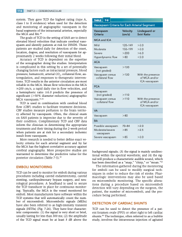
TABLE 7-5
Vasospasm Criteria for Each Arterial Segment
Vasospasm Criteria
Velocity Lindegaard or (cm/s) Sviri Ratio
MCA and ICA
Accuracy of TCD is dependent on the expertise of the sonographer doing the studies. Interpretation is complicated in this setting by a set of potentially changing factors such as intracranial pressure, blood pressure, hematocrit, arterial CO2, collateral flow, au- toregulation, and responses to therapeutic interven- tions. TCD results in the anterior circulation are most reliable in the MCA. Mean flow velocities in the MCA 200 cm/s, a rapid daily rise in flow velocities, and a hemispheric ratio 6.0 predicts the presence of significant (50% diameter reduction) angiographic MCA vasospasm.41,42
TCD is used in combination with cerebral blood flow (CBF) studies to facilitate treatment decisions. CBF studies measure perfusion to the brain territo- ry affected by vasospasm. Often, the clinical exam on SAH patients is imprecise due to the severity of their condition. Complimentary TCD and CBF data inform the clinician in determining the appropriate treatments and their timing during the 2-week period when patients are at risk for a secondary ischemic insult from vasospasm.
More research is needed to better define exact ve- locity criteria for each arterial segment and by far the MCA has the highest correlative accuracy against cerebral angiography. More prospective studies are warranted to determine the predictive value for the posterior circulation (Table 7-5).46
EMBOLI MONITORING
TCD can be used to monitor for emboli during various procedures including carotid endarterectomy, carotid stenting, cardiopulmonary bypass surgery, and neu- rological procedures. A headband is used to secure the TCD transducer in place for continuous monitor- ing. Typically, the MCA is the vessel monitored for emboli. Most manufacturers have software within the TCD systems that will automatically count the num- ber of microemboli. Microembolic signals (MESs) have also been referred to as high-intensity transient signals (HITSs) (Fig. 7-26). They have four character- istic components: (1) the signal is very short or brief, usually lasting for less than 300 ms; (2) the amplitude of the TCD signal must be at least 3 dB above the
Severe Hyperdynamic flow
200 6.0 80 3.0
Moderate 150–1993.0
ACA
Vasospasm 130 (not graded)
Vasospasm versus collateral flow
PCA
130 With the presence of MCA and/or ICA vasospasm
Vasospasm
(not graded) 110
Vasospasm versus collateral flow
VA
110 With the presence of MCA and/or ICA vasospasm
Vasospasm 80 BA
Possible vasospasm Moderate/severe
vasospasm Severe vasospasm
70–84 2.0 85 2.5
85 3.0
background signals; (3) the signal is mainly unidirec- tional within the spectral waveform; and (4) the sig- nal will produce a characteristic audible sound, which has been described as a “snap,” “chirp,” or “moan.”43
The information gathered during the monitoring for emboli can be used to modify surgical tech- niques in order to reduce the risk of stroke. Phar- macologic interventions may also be used based on microembolic monitoring. The specific altera- tions during a procedure based on microemboli detection will vary depending on the surgeon, the patient, the number of microemboli, and the pro- cedure being performed.
DETECTION OF CARDIAC SHUNTS
TCD can be used to detect the presence of a pat- ent foramen ovale (PFO) or other right to left cardiac shunts.44 The technique, often referred to as a bubble study, involves the intravenous injection of agitated
