
Page 134 - Libro 2
P. 134
114 PART 2 — CEREBROVASCULAR
artery atherosclerotic stenosis are hyperlipidemia, arterial hypertension, cigarette smoking, and dia- betes mellitus.30 Intracranial lesions may cause microemboli, which migrate into distal vascular territories causing ischemia and/or progress to sig- nificant severity or occlusion, which may result in perfusion failure, most notably in the absence of adequate collateral capacity. This latter may be a function of the site of the lesion, especially if it is located distal to the circle of Willis or due to anatomic anomalies.
Posterior Circulation
In the past, anterior circulation disease was better understood than posterior circulation. Caplan et al. developed a Posterior Circulation Registry (PCR) of patients with posterior circulation transient ischemic attacks (TIAs) or stroke. In their analysis, the inci- dence of intracranial VA stenosis was equal to that of the extracranial segments.
In the PCR, intracranial VA stenosis was present in 32% of patients, some bilaterally, and 2% had BA disease. Embolism was the most common mecha- nism of posterior circulation stroke with a prepon- derance of cardiac-origin embolism versus artery to artery. Additionally, poor outcomes were associated with cardiogenic embolism.31
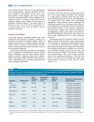
TABLE 7-3
Criteria for Intracranial Stenosis
As early as 1990, TCD criteria for stenosis and occlu- sion of the carotid siphon and MCA were reported in a large group of patients with TIA and stroke. In this seminal report by Ley-Poso and Ringelstein, 133 patients had TCD studies and conventional angiograms. Using multiple criteria, including focal velocity increases the site of stenosis, side-to-side ve- locity differences in analogous arteries, downstream hemodynamic effects in the spectral waveforms, and total lack of signal with positive identification of neighboring vessels, good results were achieved. The overall diagnostic accuracy for any lesion was 95.7%, with a sensitivity of 91.7%, and a specificity of 96.5%.32
Interpreting spectral waveforms requires knowl- edge of normal and abnormal values; appreciation of the changes in waveform morphology proximal to, at the site of, and distal to a stenosis; and a good understanding of the complex and sometimes confounding physiological variables that coexist in any individual patient at any point in time. The criteria put forth in Table 7-3 are useful as prelimi- nary measurements. There are multiple systemic factors that significantly influence velocities and need to be factored in including age, heart rate, blood pressure, hematocrit, fever, and CO2 levels (Table 7-3).33
Stenosis Criteria for the MCA (Middle Cerebral), ICA (Carotid Siphon), ACA/A1 (Anterior Cerebral), PCA/P1 (Posterior Cerebral), and Vertebral and Basilar Arteries
Mean Hemispheric
Velocity Difference Segment Depth(mm) (cm/s) (cm/s)
MCA 50–65 80–100 30 proximal
MCA distal 40–50 80–100 30 ICA (siphon) 55–65 70–90 30 ACA (A1) 65–75 70 30 PCA 56–65 50 N/A Basilar 75–110 60 N/A Vertebral 40–75 50 N/A
Ratio to
Adjacent Waveform Characteristics Artery (Poststenotic)
2 50% Turbulence, slow systolic 3 70% upstroke, covibrations 2 50% Turbulence, slow systolic 3 70% upstroke, covibrations N/A Turbulence, slow systolic
upstroke, covibrations ACA MCA Turbulence, slow systolic upstroke, covibrations PCA ACA/ICA Turbulence, slow systolic
upstroke, covibrations 30 cm/s Turbulence, slow systolic upstroke, covibrations 30 cm/s Turbulence, slow systolic
upstroke, covibrations
Velocities represent mean values. The hemispheric difference compares the same artery on the contralateral side of disease. Ratio to an adjacent artery compares the diseased artery with a normal proximal segment of the same vessel or a homologous vessel.
