
Page 145 - Libro 2
P. 145
ANKLE–BRACHIAL INDICES
The association between reduced limb systolic pres- sures (measured using plethysmography) and PAOD affecting the lower extremities was explained in the 1950s.6 The ratio of Doppler systolic pressures at bra- chial level to those at the ankle was described first in 19697 and was termed the ankle systolic pressure index. It is now known as the ankle–brachial index (ABI). Another less commonly used name for this ratio is the ankle–arm index (AAI). ABIs indicate the overall severity of PAOD between the heart and the ankle level.
An ABI is calculated by dividing the highest sys- tolic pressure at ankle level measured at either the posterior tibial artery (PTA) or the dorsalis pedis artery (DPA)/distal anterior tibial artery (ATA) by the higher of the two brachial systolic pressures. An example of an ABI calculation is:
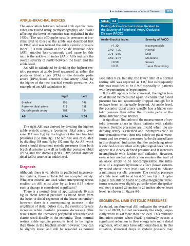
8 — Indirect Assessment of Arterial Disease 125 TABLE 8-2
Resting Ankle–Brachial Indices Related to the Severity of Peripheral Artery Occlusive Disease (PAOD)
Ankle–Brachial Index
1.30 0.901.30 0.750.89 0.500.74 0.50 0.35
Severity of PAOD
Incompressible Normal
Mild
Moderate
Severe Tissuethreatening
Brachial
Posterior tibial artery Dorsalis pedis artery
ABI
Right Left
152 146 112 158 108 154
0.74 1.04
(see Table 8-2). Initially, the lower limit of a normal resting ABI was reported as 1.0,7 but subsequently this was modified to be 0.9,10 especially in patients with hypertension or hypotension.
If the ABI appears to be abnormal, the higher bra- chial should be measured again to ensure that blood pressure has not systemically dropped enough for it to have been artifactually lowered. At ankle level, the posterior tibial artery usually has a higher sys- tolic pressure than either the dorsalis pedis or the distal anterior tibial arteries.
A significant limitation of the measurement of sys- tolic pressure arises in those patients with calcific vessels. Systolic pressures are invalid when the un- derlying artery is calcified and incompressible,11 so interpretations must then rely solely on pulse wave- forms and toe systolic pressures (to be discussed later in this chapter). Indication that the underlying artery is calcified occurs when a Doppler signal does not re- appear at a clearly defined pressure and it increases in amplitude with further cuff deflation. However, even when medial calcification renders the wall of an ankle artery to be noncompressible, the influ- ence of a negative hydrostatic effect (lower extrem- ity raised above heart level) can be used to indicate a minimum systolic pressure. The systolic pressure at ankle level will be at least 50 mm Hg if Doppler signals can still be heard or plethysmographic wave- forms remain even slightly pulsatile when the ipsilat- eral foot is raised 26 inches to 27 inches above heart level, as shown in Figure 8-3.
SEGMENTAL LIMB SYSTOLIC PRESSURES
As stated, an abnormal ABI indicates the overall se- verity of PAOD, but not necessarily the site(s), espe- cially when it is at more than one level. This multisite limitation occurs when PAOD proximally causes a significant reduction in flow energy into more distal segments, which may have additional disease. In this situation, abnormal drops in systolic pressure may
The right ABI was derived by dividing the highest ankle systolic pressure (posterior tibial artery pres- sure: 112 mm Hg) by the higher of the two brachial pressures (152 mm Hg). The left ABI was calculated by dividing 158 mm Hg by 152 mm Hg. An ABI work sheet should document systolic pressures from both brachial arteries as well as both the posterior tibial (PTA) and the dorsalis pedis (DPA)/distal anterior tibial (ATA) arteries at ankle level.
Diagnosis
Although there is variability in published interpreta- tion criteria, those in Table 8-2 are accepted widely. Whatever criteria are used when interpreting repeat studies, an ABI must alter by at least 0.15 before such a change is considered significant.8
There is a normal drop of approximately 10 mm Hg in mean arterial pressure as blood flows from the heart to distal segments of the lower extremity9; however, there is a corresponding increase in the amplitude of distal pulses (i.e., the systolic pressure increases while diastolic pressure decreases). This results from the increased peripheral resistance and elastic recoil distally in the extremity. Thus, normal resting ankle systolic pressures tend to be higher than those in the brachial artery; however, they can be slightly lower and still be regarded as normal
