
Page 146 - Libro 2
P. 146
126
PART 3 — PERIPHERAL ARTERIAL
Figure 8-3 When medial calcification renders the wall of an ankle artery to be noncompressible, the influence of a negative hydrostatic effect can be used to estimate the minimum sys- tolic pressure by raising the foot above the level of the heart.
not be exhibited in the distal segments, although differences in pulse waveforms may be discerned. To some degree, this can be remedied by measuring systolic pressures segmentally, at two or three lev- els more proximally, in addition to comparing wave- forms. The same cuffs can be used for measuring segmental systolic pressures and for volume plethys- mography studies, provided that the length of the connecting tubing does not invalidate the calibration of the plethysmograph instrument manufacturer.
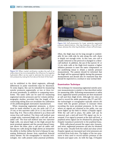
When measuring segmental pressures, a choice must be made whether to use one wide cuff (17 or 19 cm) or two narrower cuffs (10 or 12 cm) above the knee (Fig. 8-4). This is often referred to as a three-cuff versus four-cuff method. The three-cuff method uses a single wide, contoured thigh cuff, a calf cuff, and an ankle cuff. The four-cuff method uses two narrower thigh cuffs, one placed high on the thigh around the most proximal segment and the second placed lower, just above the knee, plus a calf and an ankle cuff. Having two cuffs along the thigh allows an interpreter the ability to further define the level of disease by sep- arating iliofemoral disease from superficial femoral artery disease. For the sonographer or vascular tech- nologist, two cuffs present some practical problems.
Figure 8-4 Cuff placements for lower extremity segmental pressure determinations. One leg illustrates a four-cuff tech- nique, whereas the other leg illustrates a three-cuff technique.
Often, the thigh may not be long enough to comfort- ably fit two cuffs side by side along the cuff—there is simply not enough room. In this case, one cuff is usually removed or the protocol is changed to a three- cuff method. In addition, the use of the narrow 10- or 12-cm wide cuffs at the thigh level requires a higher inflation pressure to exert the same compression on the underlying tissue to obtain a systolic pressure measurement. The patient should be informed that the thigh will be squeezed tightly during this pressure measurement and should also be reassured that this should be expected in a normal or near normal limb.
Examination Technique
The technique for measuring segmental systolic pres- sure measurements is similar to that described earlier for measuring ABIs. Following measurement at ankle level, segmental systolic pressures are then measured at calf and then thigh levels. Although two sites are used to record the ankle pressures (the PT and DP), the technologist or sonographer typically selects the vessel with the greater pressure to insonate when recording the more proximal pressures. In the case where no signals are obtained at the ankle, one can move higher up the limb to obtain a Doppler signal. With the calf cuff in place, the ankle cuff can be removed and a mid-calf level PTA signal can be in- sonated. If no signal is present at the mid-calf level, a popliteal artery signal can be obtained by placing the Doppler transducer posteriorly in the popliteal fossa.
With cuffs placed around the upper arm, forearm, and wrist, segmental pressures can also be obtained for the arms. Usually both the radial and ulnar artery Doppler signals are insonated and wrist pressures are recorded from both sites. The higher of the two pres- sures is then used to record systolic pressure at the forearm and upper arm. As with the legs, if Doppler
