
Page 148 - Libro 2
P. 148
128
PART 3 — PERIPHERAL ARTERIAL
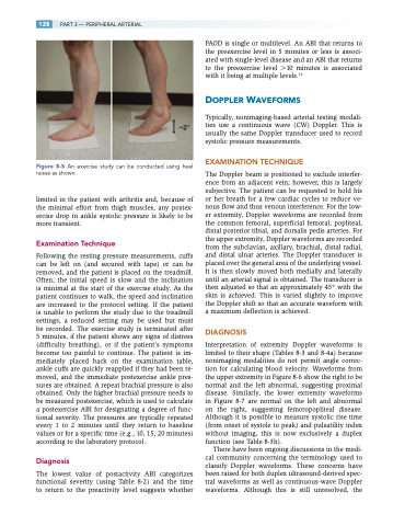
Figure 8-5 An exercise study can be conducted using heel raises as shown.
limited in the patient with arthritis and, because of the minimal effort from thigh muscles, any postex- ercise drop in ankle systolic pressure is likely to be more transient.
Examination Technique
Following the resting pressure measurements, cuffs can be left on (and secured with tape) or can be removed, and the patient is placed on the treadmill. Often, the initial speed is slow and the inclination is minimal at the start of the exercise study. As the patient continues to walk, the speed and inclination are increased to the protocol setting. If the patient is unable to perform the study due to the treadmill settings, a reduced setting may be used but must be recorded. The exercise study is terminated after 5 minutes, if the patient shows any signs of distress (difficulty breathing), or if the patient’s symptoms become too painful to continue. The patient is im- mediately placed back on the examination table, ankle cuffs are quickly reapplied if they had been re- moved, and the immediate postexercise ankle pres- sures are obtained. A repeat brachial pressure is also obtained. Only the higher brachial pressure needs to be measured postexercise, which is used to calculate a postexercise ABI for designating a degree of func- tional severity. The pressures are typically repeated every 1 to 2 minutes until they return to baseline values or for a specific time (e.g., 10, 15, 20 minutes) according to the laboratory protocol.
Diagnosis
The lowest value of postactivity ABI categorizes functional severity (using Table 8-2) and the time to return to the preactivity level suggests whether
PAOD is single or multilevel. An ABI that returns to the preexercise level in 5 minutes or less is associ- ated with single-level disease and an ABI that returns to the preexercise level 10 minutes is associated with it being at multiple levels.13
DOPPLER WAVEFORMS
Typically, nonimaging-based arterial testing modali- ties use a continuous wave (CW) Doppler. This is usually the same Doppler transducer used to record systolic pressure measurements.
EXAMINATION TECHNIQUE
The Doppler beam is positioned to exclude interfer- ence from an adjacent vein; however, this is largely subjective. The patient can be requested to hold his or her breath for a few cardiac cycles to reduce ve- nous flow and thus venous interference. For the low- er extremity, Doppler waveforms are recorded from the common femoral, superficial femoral, popliteal, distal posterior tibial, and dorsalis pedis arteries. For the upper extremity, Doppler waveforms are recorded from the subclavian, axillary, brachial, distal radial, and distal ulnar arteries. The Doppler transducer is placed over the general area of the underlying vessel. It is then slowly moved both medially and laterally until an arterial signal is obtained. The transducer is then adjusted so that an approximately 45° with the skin is achieved. This is varied slightly to improve the Doppler shift so that an accurate waveform with a maximum deflection is achieved.
DIAGNOSIS
Interpretation of extremity Doppler waveforms is limited to their shape (Tables 8-3 and 8-4a) because nonimaging modalities do not permit angle correc- tion for calculating blood velocity. Waveforms from the upper extremity in Figure 8-6 show the right to be normal and the left abnormal, suggesting proximal disease. Similarly, the lower extremity waveforms in Figure 8-7 are normal on the left and abnormal on the right, suggesting femoropopliteal disease. Although it is possible to measure systolic rise time (from onset of systole to peak) and pulsatility index without imaging, this is now exclusively a duplex function (see Table 8-3h).
There have been ongoing discussions in the medi- cal community concerning the terminology used to classify Doppler waveforms. These concerns have been raised for both duplex ultrasound-derived spec- tral waveforms as well as continuous-wave Doppler waveforms. Although this is still unresolved, the
