
Page 150 - Libro 2
P. 150
130
PART 3 — PERIPHERAL ARTERIAL
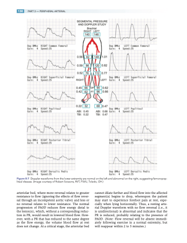
Figure 8-7 Doppler waveforms from the lower extremity are normal on the left and abnormal on the right, suggesting femoropop- liteal disease. (Image courtesy of Robert Scissons, RVT, FSVU, Toledo, OH.)
arteriolar bed, where more reversal relates to greater resistance to flow (ignoring the effects of flow rever- sal through an incompetent aortic valve) and less or no reversal relates to lower resistance. The normal progression of PAOD reduces flow energy distal to the lesion(s), which, without a corresponding reduc- tion in PR, would result in lowered blood flow. How- ever, with a PR that has reduced to the same degree as the flow energy, the volume blood flow at rest does not change. At a critical stage, the arteriolar bed
cannot dilate further and blood flow into the affected segment(s) begins to drop, whereupon the patient may start to experience forefoot pain at rest, espe- cially when lying horizontally. Thus, a resting arte- rial Doppler waveform with no flow reversal (i.e., it is unidirectional) is abnormal and indicates that the PR is reduced, probably relating to the presence of PAOD. (Note: Flow reversal will be absent immedi- ately following exercise in a normal extremity, but will reappear within 2 to 5 minutes.)
