
Page 152 - Libro 2
P. 152
132
PART 3 — PERIPHERAL ARTERIAL
A
B
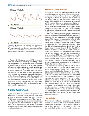
Figure 8-9 A: A normal PVR waveform from the right low thigh level exhibiting a brisk upstroke with a well-defined peak. B: The left lower thigh level PVR in this same patient illustrates moderate-to-severe PAOD changes in the waveform, causing a delayed onset to peak, which is more rounded.
Figure 8-10 illustrates normal PVR waveforms from the right lower extremity, exhibiting a brisk systolic upstroke and a concave (bows toward the baseline) shape in diastole. Each of those from the left extremity exhibit a delayed systolic upstroke and a convex diastolic component (bows away from the baseline), with reduced amplitude at the calf and an- kle level. On the left, these findings are compatible with stenosis (or occlusion with collateralization) in the iliofemoral segment, with the suggestion of additional disease at the femoropopliteal level and certainty of severe disease below the knee. Digital waveforms are also asymmetrical, with those from the left showing slight delay in the upstroke and a more rounded appearance.
DIGITAL EVALUATIONS
Digital evaluations can include both pressures and waveforms. Waveforms can be obtained either using air PVR or photoplethysmography (PPG), although PPG is most commonly performed. There are vari- ous diseases impacting the vascular system that can be diagnosed with an examination of digital blood flow. The following section will review some of the techniques employed in digital evaluations as well as specific applications.
EXAMINATION TECHNIQUE
In terms of measuring, digit pressures can be re- corded in a manner similar to other pressure mea- surements. Digital cuff diameters vary slightly but usually are 2.0 to 2.5 cm. Typically, PPG is the most convenient modality for measuring digital pres- sures, although PVR is an alternative. The use of a PVR requires Doppler to insonate the digital ar- teries. It is not easy to maintain the Doppler ultra- sound beam within the lumen of a digital artery (which is typically only 1 to 2 mm), so using PVRs is a less convenient method for measuring digital systolic pressures.
PPGs are not true plethysmographic instruments because they cannot be calibrated in volume terms; however, they are convenient for recording arterial pulses. They operate by transmitting infrared light into tissue and detecting variations in the light re- flected from underlying blood flow, from a depth of 1 to 3 mm. They are maintained in contact with the skin with double-sided tape (Fig. 8-11A), with a Velcro strap, or with a clip-style device (Fig. 8-11B). The PPG is placed on the digit and, after a brief pe- riod of 1 to 2 seconds, the sensor on the unit detects the reflected infrared light, which is then displayed as a waveform. PPG and PVR instruments provide comparably shaped waveforms and so are inter- preted similarly, with normal characteristics being a brisk systolic upstroke, a well-defined peak, and a concave shape as the shape returns to the baseline during diastole (see Fig. 8-9A).
In addition to recording arterial pulses, PPGs can also be used to measure digital systolic pres- sures. The pulses are recorded using a slow chart or sweep speed and adjusting the gain to give an am- plitude approximately one-third of the chart width (Fig. 8-12). Limb or digit movement can intrude to confuse the point at which the pulses return if the recording has greater amplitude. The cuff is inflat- ed until pulsatility is abolished, then deflated until pulses are seen. Hands or feet can be warmed to increase pulse amplitude. This can be accomplished by heating a small towel in a microwave for 1 min- ute. The towel can be wrapped around each hand or foot while the history is being documented. Having the recording with an enhanced pulse amplitude en- ables the point of pulse return to be determined with greater confidence.
DIAGNOSIS
A toe pressure can be used to calculate a toe–brachial index (TBI), which should be 0.8 to be reported as normal (see Table 8-3e).16 This is similar to measur- ing an ankle pressure for calculating an ABI. TBIs are particularly useful when the ankle vessels are
