
Page 147 - Libro 2
P. 147
signals are not audible distally (at the wrist level), the Doppler transducer can be placed more proxi- mally over the brachial artery to record the pressure in the upper arm cuff.
Diagnosis
Table 8-3 lists interpretation criteria for the lower extremity. Systolic pressures usually increase as blood flows distally along the lower extremity, al- though there can also be a slight drop (Table 8-3d). However, any reduction in distal pressure should be 30 mm Hg between adjacent segments (thigh level to calf, calf to ankle) (see Table 8-3i)12 with drops greater than this being associated with the presence of proximal obstruction.
The width of the thigh cuff(s) changes the criteria when interpreting thigh pressure(s). Normal systol- ic pressure measured from a single large thigh cuff (17 or 19 cm width) should be equal to the higher of the two brachial pressures (see Table 8-3f). Because narrower cuffs (10 or 12 cm width) require a higher inflation pressure, the normal systolic pressure at high thigh level (using a narrow cuff) needs to be 30 mm Hg or so above the higher brachial pressure (see Table 8-3g).
PAOD is less common in the upper extremity com- pared with the lower but, when present, it is found most likely in the subclavian and proximal axillary arteries. Table 8-4 lists interpretation criteria for the upper extremity. A 75% diameter reduction in ei- ther of these arteries will cause a 20 mm Hg differ- ence between brachial systolic pressures. Typically, segmental pressures are accompanied by abnormal
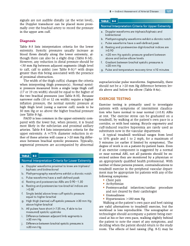
TABLE 8-3
Normal Interpretation Criteria for Lower Extremity
a. Doppler waveforms proximal to knee are triphasic/ biphasic and bidirectional
b. Plethysmographywaveformsexhibitadicroticnotch
c. Pulse waveforms have a well-defined peak
d. Resting and postexercise ABIs are 0.901.30
e. Resting and postexercise toe-brachial indices are
0.80
f. Single (wide) above knee cuff systolic pressure
equal to higher brachial
g. Highthigh(narrow)cuffsystolicpressure30mmHg
above higher brachial
h. All pulses have short (135 ms, if able to be
measured) systolic upstroke
i. Difference between adjacent limb segments is
30 mm Hg
j. Difference between brachial systolic pressures is 20 mm Hg
8 — Indirect Assessment of Arterial Disease 127 TABLE 8-4
Normal Interpretation Criteria for Upper Extremity
a. Doppler waveforms are triphasic/biphasic and bidirectional
b. Plethysmographywaveformsexhibitadicroticnotch
c. Pulse waveforms have a well-defined peak
d. Resting and postexercise digit-brachial indices are
0.90
e. 20 mm Hg systolic pressure gradient between
above and below elbow levels
f. Gradient between brachial systolic pressures is
20 mm Hg
g. Pulse and temperature recovery time 10 minutes
supraclavicular pulse waveforms. Segmentally, there should not be a 20 mm Hg difference between lev- els above and below the elbow (Table 8-4e).
EXERCISE TESTING
Exercise testing is primarily used to investigate patients with symptoms of intermittent claudica- tion who have normal to near normal (0.8) ABIs at rest. The exercise stress can be graduated on a treadmill, by walking at the patient’s own pace in a corridor, or with heel raising. Bicycle ergometry and cuff-induced reactive hyperemia are rarely used as substitutes now in the vascular department.
A typical treadmill workload ranges from level to 10% grade and 1 to 2 mph, for a maximum of 5 minutes (or earlier if limited by symptoms). The degree of work is on a patient-by-patient basis. Even if an exercise component is suggested by a normal or near normal ABI, not all patients should be ex- ercised unless they are monitored by a physician or an appropriately qualified health professional. With neither of these persons present, contraindications to treadmill exercise in the peripheral vascular depart- ment may be appropriate for patients with any of the following symptoms:
• Chest pain
• Arrhythmias
• Postmyocardial infarction/cardiac procedure
and not cleared by their cardiologist
• Unsteadiness
• Hypertension 180 mm Hg
Walking at the patient’s own pace and heel raising
are valid alternatives to treadmill exercise, but the workload is less reproducible. The sonographer or technologist should accompany a patient being exer- cised at his or her own pace, walking slightly behind the patient to note the onset of any symptoms, and deciding when the patient should return to the study room. The effects of heel raising (Fig. 8-5) may be
