
Page 191 - Libro 2
P. 191
11 — Ultrasound Assessment of Arterial Bypass Grafts
171
AB
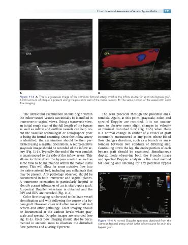
Figure 11-3 A: This is a grayscale image of the common femoral artery, which is the inflow source for an in situ bypass graft. A mild amount of plaque is present along the posterior wall of the vessel (arrow). B: The same portion of the vessel with color flow imaging.
The ultrasound examination should begin within the inflow vessel. Vessels can initially be identified in transverse or sagittal views. Using a transverse view, an initial rough scan of the full length of the bypass as well as inflow and outflow vessels can help ori- ent the vascular technologist or sonographer prior to being the formal scanning. Once the inflow artery is identified, the examination should be then per- formed using a sagittal orientation. A representative grayscale image should be recorded of the inflow ar- tery (Fig. 11-3). Typically, the end of the vein conduit is anastomosed to the side of the inflow artery. This allows for flow down the bypass conduit as well as some flow to be maintained within the native distal artery. This will allow for some nutritive flow into the native arterial bed, including any collaterals that may be present. Any pathology observed should be documented in both transverse and sagittal planes. A transverse orientation is particularly helpful to identify patent tributaries of an in situ bypass graft. A spectral Doppler waveform is obtained and the PSV and EDV are recorded (Fig. 11-4).
Color flow imaging can be used to facilitate vessel identification and with following the course of a by- pass graft. However, color will often mask small wall defects and other pathology. Color imaging should be documented at the various levels where gray- scale and spectral Doppler images are recorded (see Fig. 11-3). Color flow imaging should also be docu- mented in stenotic areas to illustrate the disturbed flow patterns and aliasing if present.
The scan proceeds through the proximal anas- tomosis. Again, at this point, grayscale, color, and spectral Doppler are recorded. It is not uncom- mon to observe some slight changes in velocity or minimal disturbed flow (Fig. 11-5) when there is a normal change in caliber of a vessel or graft commonly encountered at any point where blood flow changes direction, such as a branch or anas- tomosis between two conduits of differing size. Continuing down the leg, the entire portion of each bypass graft should be examined. Simultaneous duplex mode observing both the B-mode image and spectral Doppler analysis is the ideal method for looking and listening for any potential bypass
Figure 11-4 A normal Doppler spectrum obtained from the common femoral artery, which is the inflow source for an in situ bypass graft.
