
Page 192 - Libro 2
P. 192
172
PART 3 — PERIPHERAL ARTERIAL
Figure 11-5 A Doppler spectrum taken at the proximal anas- tomosis of a bypass graft. Slight turbulence is present.
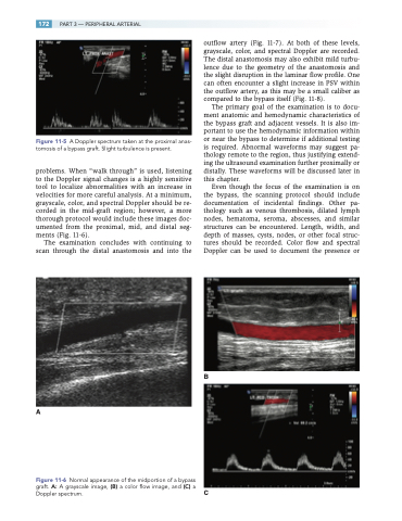
problems. When “walk through” is used, listening to the Doppler signal changes is a highly sensitive tool to localize abnormalities with an increase in velocities for more careful analysis. At a minimum, grayscale, color, and spectral Doppler should be re- corded in the mid-graft region; however, a more thorough protocol would include these images doc- umented from the proximal, mid, and distal seg- ments (Fig. 11-6).
The examination concludes with continuing to scan through the distal anastomosis and into the
A
outflow artery (Fig. 11-7). At both of these levels, grayscale, color, and spectral Doppler are recorded. The distal anastomosis may also exhibit mild turbu- lence due to the geometry of the anastomosis and the slight disruption in the laminar flow profile. One can often encounter a slight increase in PSV within the outflow artery, as this may be a small caliber as compared to the bypass itself (Fig. 11-8).
The primary goal of the examination is to docu- ment anatomic and hemodynamic characteristics of the bypass graft and adjacent vessels. It is also im- portant to use the hemodynamic information within or near the bypass to determine if additional testing is required. Abnormal waveforms may suggest pa- thology remote to the region, thus justifying extend- ing the ultrasound examination further proximally or distally. These waveforms will be discussed later in this chapter.
Even though the focus of the examination is on the bypass, the scanning protocol should include documentation of incidental findings. Other pa- thology such as venous thrombosis, dilated lymph nodes, hematoma, seroma, abscesses, and similar structures can be encountered. Length, width, and depth of masses, cysts, nodes, or other focal struc- tures should be recorded. Color flow and spectral Doppler can be used to document the presence or
B
C
Figure 11-6 Normal appearance of the midportion of a bypass graft. A: A grayscale image, (B) a color flow image, and (C) a Doppler spectrum.
