
Page 194 - Libro 2
P. 194
174
PART 3 — PERIPHERAL ARTERIAL
AB
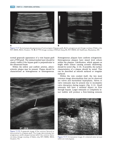
Figure 11-9 Normal grayscale appearance of various types of bypass grafts. A: An autogenous vein (Image courtesy of Debra Joly, RVT, RDMS, RDCS, Houston, TX). B: A PTFE (Image courtesy of William Zang, BS, RVT, RDMS, GE Healthcare, Wauwatosa, WI).
normal grayscale appearance of a vein bypass graft and a PTFE graft. The intimal-medial layer should be clearly visible if the bypass graft is perpendicular to the ultrasound beam.
Within the inflow and outflow arteries, athero- sclerotic plaque may be present. Plaque should be characterized as homogeneous or heterogeneous.
Figure 11-10 A grayscale image of the common femoral ar- tery proximal to a bypass graft. The image demonstrates het- erogeneous calcific plaque (arrow) within the common femo- ral artery. (Image courtesy of Debra Joly, RVT, RDMS, RDCS, Houston, TX.)
Homogeneous plaques have uniform echogenicity. Heterogeneous plaques have mixed level echoes within the plaques. Calcification, which appears as bright white echoes causing acoustic shadowing, should be noted (Fig. 11-10). If possible, the surface characteristics of a plaque should be noted. This can be described as smooth surfaced or irregular surfaced.
Within the vein conduit itself, the two most common image abnormalities that can be observed are valves and myointimal hyperplasia. Valves or valve remnants may be present due to incomplete valve disruption during surgery (Fig. 11-11). Small remnants will have a minimal impact on flow through bypass. Larger remnants or completely in- tact leaflets will produce a flow-limiting stenosis
Figure 11-11 A transverse image of a retained valve (arrows) within an in situ bypass.
