
Page 196 - Libro 2
P. 196
176
PART 3 — PERIPHERAL ARTERIAL
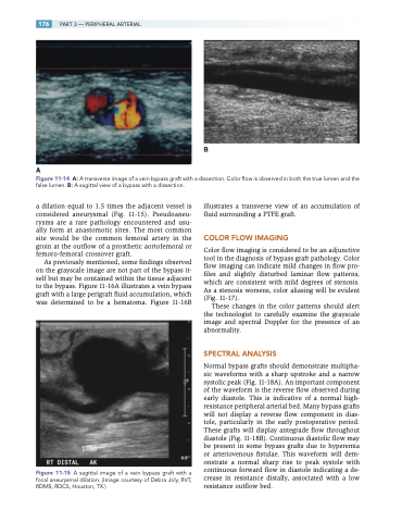
A
Figure 11-14 A: A transverse image of a vein bypass graft with a dissection. Color flow is observed in both the true lumen and the false lumen. B: A sagittal view of a bypass with a dissection.
a dilation equal to 1.5 times the adjacent vessel is considered aneurysmal (Fig. 11-15). Pseudoaneu- rysms are a rare pathology encountered and usu- ally form at anastomotic sites. The most common site would be the common femoral artery in the groin at the outflow of a prosthetic aortofemoral or femoro-femoral crossover graft.
As previously mentioned, some findings observed on the grayscale image are not part of the bypass it- self but may be contained within the tissue adjacent to the bypass. Figure 11-16A illustrates a vein bypass graft with a large perigraft fluid accumulation, which was determined to be a hematoma. Figure 11-16B
Figure 11-15 A sagittal image of a vein bypass graft with a focal aneurysmal dilation. (Image courtesy of Debra Joly, RVT, RDMS, RDCS, Houston, TX.)
illustrates a transverse view of an accumulation of fluid surrounding a PTFE graft.
COLOR FLOW IMAGING
Color flow imaging is considered to be an adjunctive tool in the diagnosis of bypass graft pathology. Color flow imaging can indicate mild changes in flow pro- files and slightly disturbed laminar flow patterns, which are consistent with mild degrees of stenosis. As a stenosis worsens, color aliasing will be evident (Fig. 11-17).
These changes in the color patterns should alert the technologist to carefully examine the grayscale image and spectral Doppler for the presence of an abnormality.
SPECTRAL ANALYSIS
Normal bypass grafts should demonstrate multipha- sic waveforms with a sharp upstroke and a narrow systolic peak (Fig. 11-18A). An important component of the waveform is the reverse flow observed during early diastole. This is indicative of a normal high- resistance peripheral arterial bed. Many bypass grafts will not display a reverse flow component in dias- tole, particularly in the early postoperative period. These grafts will display antegrade flow throughout diastole (Fig. 11-18B). Continuous diastolic flow may be present in some bypass grafts due to hyperemia or arteriovenous fistulae. This waveform will dem- onstrate a normal sharp rise to peak systole with continuous forward flow in diastole indicating a de- crease in resistance distally, associated with a low resistance outflow bed.
B
