
Page 195 - Libro 2
P. 195
11 — Ultrasound Assessment of Arterial Bypass Grafts
175
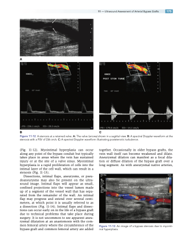
A
BC
Figure 11-12 A stenosis at a retained valve. A: The valve (arrows) shown in a sagittal view. B: A spectral Doppler waveform at the stenosis with a PSV of 236 cm/s. C: A spectral Doppler waveform illustrating poststenotic turbulence.
(Fig. 11-12). Myointimal hyperplasia can occur along any point of the bypass conduit but typically takes place in areas where the vein has sustained injury or at the site of a valve sinus. Myointimal hyperplasia is a rapid proliferation of cells into the intimal layer of the cell wall, which can result in a stenosis (Fig. 11-13).
Dissections, intimal flaps, aneurysms, or pseu- doaneurysms may also be present on the ultra- sound image. Intimal flaps will appear as small, confined projections into the vessel lumen made up of a segment of the vessel wall that has sepa- rated from the remainder of the wall. An intimal flap may progress and extend over several centi- meters, at which point it is usually referred to as a dissection (Fig. 11-14). Intimal flaps and dissec- tions can occur early on in the life of a bypass graft due to technical problems that take place during surgery. It is not uncommon to see apparent aneu- rysmal dilatation at an anastomosis with the com- mon femoral artery where the circumference of the bypass graft and common femoral artery are added
together. Occasionally in older bypass grafts, the vein wall itself can become weakened and dilate. Aneurysmal dilation can manifest as a focal dila- tion or diffuse dilation of the bypass graft over a long segment. As with aneurysmal native arteries,
Figure 11-13 An image of a bypass stenosis due to myointi- mal hyperplasia.
