
Page 217 - Libro 2
P. 217
13 — Special Considerations in Evaluating Nonatherosclerotic Arterial Pathology
197
AB
CD
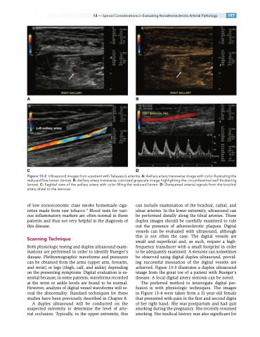
Figure 13-2 Ultrasound images from a patient with Takayasu’s arteritis. A: Axillary artery transverse image with color illustrating the reduced flow lumen (arrow). B: Axillary artery transverse colorized grayscale image highlighting the circumferential wall thickening (arrow). C: Sagittal view of the axillary artery with color filling the reduced lumen. D: Dampened arterial signals from the brachial artery distal to the stenosis.
of low socioeconomic class smoke homemade ciga- rettes made from raw tobacco.9 Blood tests for vari- ous inflammatory markers are often normal in these patients and thus not very helpful in the diagnosis of this disease.
Scanning Technique
Both physiologic testing and duplex ultrasound exam- inations are performed in order to identify Buerger’s disease. Plethysmographic waveforms and pressures can be obtained from the arms (upper arm, forearm, and wrist) or legs (thigh, calf, and ankle) depending on the presenting symptoms. Digital evaluation is es- sential because, in some patients, waveforms recorded at the wrist or ankle levels are found to be normal. However, analysis of digital vessel waveforms will re- veal the abnormality. Standard techniques for these studies have been previously described in Chapter 8.
A duplex ultrasound will be conducted on the suspected extremity to determine the level of arte- rial occlusion. Typically, in the upper extremity, this
can include examination of the brachial, radial, and ulnar arteries. In the lower extremity, ultrasound can be performed distally along the tibial arteries. These duplex images should be carefully examined to rule out the presence of atherosclerotic plaques. Digital vessels can be evaluated with ultrasound, although this is not often the case. The digital vessels are small and superficial and, as such, require a high- frequency transducer with a small footprint in order to be adequately examined. A stenosis can sometimes be observed using digital duplex ultrasound, provid- ing successful insonation of the digital vessels are achieved. Figure 13-3 illustrates a duplex ultrasound image from the great toe of a patient with Buerger’s disease. A focal digital artery stenosis can be noted.
The preferred method to interrogate digital per- fusion is with physiologic techniques. The images in Figure 13-4 were taken from a 31-year-old female that presented with pain in the first and second digits of her right hand. She was postpartum and had quit smoking during the pregnancy. She recently resumed smoking. Her medical history was also significant for
