
Page 218 - Libro 2
P. 218
198
PART 3 — PERIPHERAL ARTERIAL
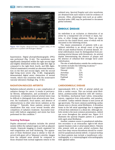
Figure 13-3 Duplex ultrasound from a digital artery of the great toe in a patient with Buerger’s disease.
hypertension. Digital photoplethysmography (PPG) was performed (Fig. 13-4A). The waveforms were significantly dampened within the right second digit and slightly diminished within the right first digit as compared to the right third, fourth, and fifth digits. Clinical examination revealed an abnormal demarca- tion on the first and second digits, with the second digit being more severe (Fig. 13-4B). Angiography demonstrated digital artery obstruction of several vessels, which was consistent with the noninvasive vascular laboratory findings (Fig. 13-4C).
RADIATION-INDUCED ARTERITIS
Radiation-induced arteritis is a rare complication of radiation therapy for cancer. It results in perivascu- lar fibrosis, inflammation, and acceleration of ath- erosclerosis. A radiation-induced arterial lesion may be difficult to distinguish from atherosclerosis. How- ever, the localization, focal nature, and absence of atherosclerosis in other sites favors radiation as the etiology.10,11 Typically, these patients present with claudication that may occur several months after completion of radiation treatment. Successful treat- ment with balloon angioplasty and stents has been performed for this condition.12
Scanning Technique
Duplex ultrasound evaluation includes the arterial system within the radiated region as well as adjacent vessels. Grayscale imaging is closely examined for wall irregularities and wall thickening. The appear- ance of these thickened areas is similar to that ob- served with giant cell or Takayasu’s arteritis. Images from the radiated areas should be compared to those obtained from normal segments remote to the
radiated area. Spectral Doppler and color waveforms are obtained from each vessel to look for evidence of stenosis. Often, physiologic tests such as an ankle– brachial index (ABI) may be performed to document global ischemia.
EMBOLIC DISEASE
An embolism is an occlusion or obstruction of an artery by a transported clot of blood or mass, bac- teria, or other foreign substance. There are many sources for the clotted blood, some of which will be discussed in the following section.
The classic presentation of patients with a pe- ripheral embolism is an abrupt onset of leg pain with no past medical history of arterial disease. Ar- terial embolization must be distinguished from pre- existing arterial disease with thrombosis. An arterial duplex demonstrating the absence of plaque and the absence of collateral flow strongly favor acute embolization.
The site of embolization outside the cerebrovascu- lar system includes the following locations:13
Upper extremity 14% Visceral 7% Aortoiliac 22% Femoral 36% Popliteal 15% Other 6%
CARDIOEMBOLIC DISEASE
Approximately 80% to 99% of arterial emboli are from a cardiac source. This can include atrial fibril- lation, postmyocardial infarction with left ventricu- lar thrombus, mechanical heart valves, intracardiac tumors, vegetation from endocarditis, and paradoxi- cal emboli. Cardiac embolic disease is seen across all age spectrums. The most common underlying cardiac disease seen is chronic atrial fibrillation. A thrombus forms in the left atrial appendage due to stagnation of blood then embolizes to a distal site. Although em- bolic stroke is the most common presentation, emboli can occur throughout the arterial tree. Figure 13-5 illustrates the spectral Doppler pattern in a patient with rapid atrial fibrillation.
Another common source is paradoxical emboliza- tion secondary to an intracardiac right-to-left shunt (patent foramen ovale or atrial septal defect). Patients who present with an acute arterial occlusion that may have deep venous thrombosis should be consid- ered for paradoxical arterial emboli. A typical workup for these patients may include a transthoracic echo- cardiography or a transesophageal echocardiography with agitated saline.
