
Page 220 - Libro 2
P. 220
200
PART 3 — PERIPHERAL ARTERIAL
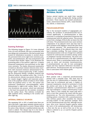
Figure 13-5 Doppler spectrum of a patient with atrial fibrillation.
Scanning Technique
The following images in Figure 13-6 were obtained from a 26-year-old female. She was a nonsmoker who was taking oral contraceptives. She had recently been treated for dehydration secondary to alcohol abuse. She presented with severe right lower extremity pain, pallor, and pulselessness. She was normotensive and in normal sinus rhythm. Figure 13-6A illustrates the presenting pallor of the patient’s right foot. A duplex ultrasound examination was performed on the right lower extremity. The duplex ultrasound examination revealed an abnormal common femoral artery wave- form with increased resistance demonstrated by the abrupt “spike” noted in systole (Fig. 13-6B). Continu- ing the ultrasound distally, echogenic material was observed within the popliteal artery (Fig. 13-6C,D). No atherosclerotic disease was present within any of the vessels imaged. Sagittal and transverse projec- tions of the popliteal vessels revealed no flow within either the popliteal artery or vein. The patient also underwent transthoracic echocardiography, which demonstrated a right-to-left cardiac shunt. A deep vein thrombosis was present, which had embolized to the heart but passed into the systemic circulation via the cardiac shunt. This embolus occluded the right popliteal artery, resulting in the acutely isch- emic right leg.
ARTERIAL EMBOLIC SOURCES
The remaining 10% to 20% of emboli arise from out- side of the heart. Large upper extremity arteries such as the subclavian arteries may be a source. Disease within the aorta or iliac, femoral, or popliteal arteries may also result in emboli. This disease may be in the form of ulcerated plaques or mural thrombus aneurysms.
TRAUMATIC AND IATROGENIC ARTERIAL INJURY
Various arterial injuries can result from vascular trauma or can result iatrogenically during another procedure. These injuries can include pseudoaneu- rysms, arteriovenous fistulae (AVF), vessel thrombo- ses, emboli, or dissections.
PSEUDOANEURYSMS
Due to the increased numbers of angiographic pro- cedures, the incidence of pseudoaneurysms has in- creased significantly. A pseudoaneurysm or false aneurysm is a pulsating encapsulated hematoma that communicates with the adjacent artery. This process occurs from a leakage of blood after an injury into the soft tissue. The pseudoaneurysm may cause extrinsic compression of an adjacent nerve and may lead to nerve irritation with tingling or shock-like pain down the affected extremity. The pseudoaneurysm may lead to compression of the adjacent deep vein, lead- ing to extremity swelling. The most common cause of pseudoaneurysms is the use of large-bore catheters required for endovascular procedures during cardiac or peripheral vascular interventions. The most com- mon site for a pseudoaneurysm is the right common femoral artery. Blunt or penetrating trauma may also cause an injury and secondary pseudoaneurysm. A pseudoaneurysm can also be seen postsurgical bypass and may represent dehiscence secondary to graft infection. Pseudoaneurysms also are frequently seen in association with dialysis grafts.
Scanning Technique
Most patients with a suspected pseudoaneurysm present with a pulsatile mass. This mass may be over the site of a catheterization or traumatic injury. There may be ecchymosis and pain. A duplex ul- trasound examination should be performed of both the arteries and veins in the region. Figure 13-7A illustrates a pseudoaneurysm observed in a patient who had undergone a cardiac catheterization 4 days prior. There is color outside the vessel wall with a neck or tract that connects the vessel with the pseu- doaneurysm sac. The color demonstrates a red-blue swirling pattern consistent with a pseudoaneurysm. This red-blue pattern often resembles the Chinese yin-yang symbol. In this particular example, there are two pseudoaneurysm sacs that, although uncom- mon, does occur. Spectral Doppler patterns within the neck of the pseudoaneurysm will demonstrate a flow into the sac as well as flow out of the sac, which is referred to as a to-and-fro Doppler pattern (Fig. 13-7B).
