
Page 221 - Libro 2
P. 221
13 — Special Considerations in Evaluating Nonatherosclerotic Arterial Pathology
201
AB
CD
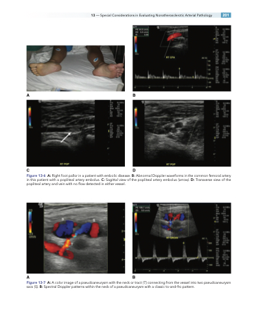
Figure 13-6 A: Right foot pallor in a patient with embolic disease. B: Abnormal Doppler waveforms in the common femoral artery in this patient with a popliteal artery embolus. C: Sagittal view of the popliteal artery embolus (arrow). D: Transverse view of the popliteal artery and vein with no flow detected in either vessel.
AB
Figure 13-7 A: A color image of a pseudoaneurysm with the neck or tract (T) connecting from the vessel into two pseudoaneurysm sacs (S). B: Spectral Doppler patterns within the neck of a pseudoaneurysm with a classic to-and-fro pattern.
