
Page 222 - Libro 2
P. 222
202
PART 3 — PERIPHERAL ARTERIAL
ARTERIOVENOUS FISTULAE
Traumatic AVF can occur following arterial trauma such as a direct stab wound, gunshot wound, or blunt trauma. Most iatrogenic AVFs result as a complication of percutaneous femoral artery cath- eterization. Central venous catheterization occa- sionally leads to AVFs and rarely, AVFs are seen after total knee replacement or lumbosacral sur- gery. Patients with an AVF usually present with signs and symptoms at the site of intervention or injury. Typically, a new bruit is ascultated, a thrill may be palpated, or a hematoma is present. The patients are often referred for an ultrasound to rule out the presence of a pseudoaneurysm. Rarely, both a pseudoaneurysm and AVF may be present at the same location.
Scanning Technique
The gold standard to detect an AVF remains digi- tal subtraction angiography, but duplex ultrasound is a frequently used alternative for detection of
A
C
an AVF. The characteristic findings on ultrasound include:
• High diastolic flow in an artery proximal to AVF
• High velocity turbulent flow (arterialized venous
flow) in the vein near the fistula connection
• A color bruit may be seen at the site near the
fistula connection
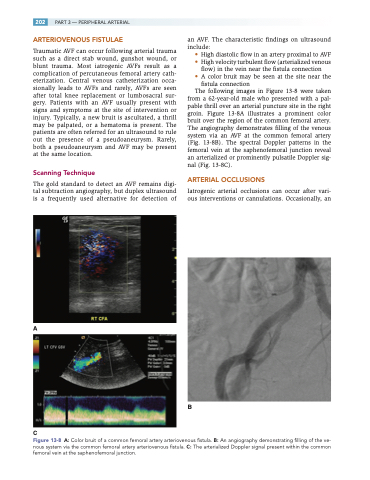
The following images in Figure 13-8 were taken
from a 62-year-old male who presented with a pal- pable thrill over an arterial puncture site in the right groin. Figure 13-8A illustrates a prominent color bruit over the region of the common femoral artery. The angiography demonstrates filling of the venous system via an AVF at the common femoral artery (Fig. 13-8B). The spectral Doppler patterns in the femoral vein at the saphenofemoral junction reveal an arterialized or prominently pulsatile Doppler sig- nal (Fig. 13-8C).
ARTERIAL OCCLUSIONS
Iatrogenic arterial occlusions can occur after vari- ous interventions or cannulations. Occasionally, an
B
Figure 13-8 A: Color bruit of a common femoral artery arteriovenous fistula. B: An angiography demonstrating filling of the ve- nous system via the common femoral artery arteriovenous fistula. C: The arterialized Doppler signal present within the common femoral vein at the saphenofemoral junction.
