
Page 224 - Libro 2
P. 224
204
PART 3 — PERIPHERAL ARTERIAL
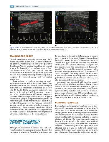
AB
Figure 13-10 A: The left popliteal artery in a patient with popliteal entrapment. With the leg in a relaxed neutral position, the PSV is 84 cm/s. B: With plantar flexion, the popliteal artery velocities increase to 130 cm/s.
SCANNING TECHNIQUE
Clinical examination typically reveals that distal pulses are present at rest with the ankle in the neu- tral position. Pulses disappear with active plantar or dorsiflexion. Various imaging modalities can be used to aid in the diagnosis of popliteal entrapment. How- ever, interpretation of the lower extremity vascular examination must relate to the patient’s symptoms because many asymptomatic patients will partially compress the popliteal artery with provocative maneuvers.
Ultrasound can be employed to image the popli- teal artery at rest and with active plantar flexion. Nor- mal velocities at rest will become altered during the maneuver and demonstrate diminished or no flow (Fig. 13-10A,B). Digital subtraction angiography can also be performed and will demonstrate compres- sion of the popliteal artery with active maneuvers. However, other imaging modalities such as magnetic resonance imaging or computerized tomography are often employed. These other modalities can not only provide information about the vascular system, but also can identify the skeletal/muscular features of the popliteal fossa. These anatomic details can identify those components responsible for the compression of the vessel. It can also exclude other pathology such as adventitial cystic disease of the popliteal artery.
NONATHEROSCLEROTIC ARTERIAL ANEURYSMS
Although atherosclerosis is a major cause of aneu- rysm formation, there are multiple other diseases that can result in aneurysm formation. Aneurysms can
be associated with various inflammatory processes such as some of the arteritis diseases discussed ear- lier in this chapter. Takayasu’s disease involves large arteries and typically causes flow-reducing stenosis of the aortic arch arteries. However, aneurysms are the most frequent fatal complication of Takayasu’s arteritis. The reported incidence of aneurysms varies with one recent paper reporting a 45% occurrence of aortic aneurysms in these patients.15 Other rare in- flammatory diseases, including Behcet’s syndrome, polyarteritis nodosa, and Kawasaki’s disease, can also be associated with aneurysms.
Aneurysms are associated with inherited matrix defects such as Marfan’s syndrome. Marfan’s syn- drome is a connective tissue disorder that is typically associated with aortic arch aneurysms. Ehlers-Danlos syndrome (EDS) is another well-known and often sus- pected cause of aneurysms. EDS patients have a con- genital defect in type III collagen and this can lead to arterial rupture with or without an aneurysm.
SCANNING TECHNIQUE
Duplex ultrasound imaging has long been used to iden- tify arterial aneurysms. Aneurysms of the aortic arch and thoracic aorta are best identified with various types of angiography. When evaluating peripheral arteries for aneurysm formation, measurements of vessel diameter should be obtained both proximal and distal to an area of suspected dilation. If a vessel diameter increases at least 50% as compared to the native, more proximal adjacent segment of artery, this vessel is considered an- eurysmal. The following images in Figure 13-11 were obtained from a 51-year-old male with Marfan’s syn- drome. He had previously undergone an aortic arch and valve repair. He presented with left lower leg pain,
