
Page 225 - Libro 2
P. 225
13 — Special Considerations in Evaluating Nonatherosclerotic Arterial Pathology
205
AB
C
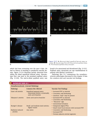
which had been worsening over the past 4 days. He had no history of smoking or peripheral vascular dis- ease. Figure 13-11A illustrates grossly abnormal flow within the distal superficial femoral artery. Staccato- type flow was seen in the proximal popliteal artery (Fig. 13-11B). The mid-to-distal popliteal artery was
Figure 13-11 A: Abnormal distal superficial femoral artery ve- locities. B: Staccato-type flow with the proximal popliteal artery. C: Thrombosed popliteal artery aneurysm.
found to be aneurysmal and thrombosed (Fig. 13-11C). Popliteal artery aneurysms are rare manifestations in patients with Marfan’s syndrome.16
Pathology Box 13-1 summarizes the nonathero- sclerotic pathologies discussed in this chapter. It lists the common sites and vascular test findings.
PATHOLOGY BOX 13-1
Nonatherosclerotic Arterial Pathology
Pathology Common Site Affected Vascular Test Findings
Giant cell arteritis
Takayasu’s arteritis
Buerger’s disease
Radiation-induced arteritis
Superficial temporal artery; extracranial arteries; occasionally, aortic arch branches
Aortic arch and its branches
Small- and medium-sized arteries; digital vessels
Any artery
• Increased PSV at stenosis
• Concentric wall thickening
• Anechoic “halo” may be present
• Increased PSV at stenosis
• Concentric wall thickening
• “Macaroni” sign
• Aneurysmal formation
• Dampened digital PPGs
• Small vessel focal stenosis with increased PSV • Concentric wall thickening
• Increased PSV
• Normal adjacent arterial segments
