
Page 226 - Libro 2
P. 226
206
PART 3 — PERIPHERAL ARTERIAL
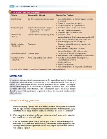
PATHOLOGY BOX 13-1 (continued)
Pathology Common Site Affected Vascular Test Findings
Embolic disease
Pseudoaneurysm
Arteriovenous fistula
Arterial occlusion/ thrombosis
Popliteal entrapment
Nonatherosclerotic aneurysm
Cerebrovascular vessels; any artery
Common femoral artery; dialysis fistulae/grafts; bypass graft anastomoses
Common femoral artery; any artery
Any artery
Popliteal artery
Aorta; large and medium arteries
• Increased resistance in Doppler signals proximal to embolus
• Echogenic material within vessel
• No atherosclerosis in adjacent vessel
• Color flow outside normal vessel walls
• Yin-yang swirling color pattern
• To-and-fro signal in neck or tract
• Color tissue bruit
• Increased diastolic flow in artery proximal to AVF • High velocity, turbulent signals at fistula site
• Prominently pulsatile venous signals
• Echogenic material in vessel at puncture site
• Poor color filling
• Increased PSV with partial occlusion
• Increased resistance with total occlusion
• PSV normal in neutral, resting position
• Increased PSV with active plantar or dorsiflexion • Diameter increase of 50% as compared to
adjacent proximal vessel
• No atherosclerosis
• May contain thrombus
• Increased resistance if completely thrombosed
PSV, peak systolic velocity; PPG, photoplethysmography; AVF, arteriovenous fistula.
SUMMARY
In summary, the majority of patients presenting for a peripheral arterial ultrasound examination will likely have atherosclerosis as the disease identified. There are nu- merous other pathologies that will produce peripheral arterial disease. It is impor- tant for the sonographer or vascular technologist to be familiar with these entities and their ultrasound characteristics. These uncommon causes of arterial disease should be suspected, particularly in patients without the standard risk factors for atherosclerosis.
Critical Thinking Questions
1. You are examining a patient with a 35 mm Hg brachial blood pressure difference, with the right brachial blood pressure lower than the left. Will the spectral analy- sis or the B-mode image be more helpful to determine the cause of a stenosis?
2. When evaluating a patient for Buerger’s disease, which noninvasive vascular tests would you perform and why?
3. What are three iatrogenic arterial pathologies that can arise following cath- eterization of the common femoral artery for coronary artery angioplasty and stenting? Which of these problems would most impact the common femoral vein Doppler signals?
