
Page 231 - Libro 2
P. 231
14 — Duplex Imaging of the Lower Extremity Venous System 211
cause of preventable death in hospital mortality in the United States. It has been estimated that, in the United States, there are greater than 500,000 cases of DVT each year with more than 50% of those cases going unrecognized. There are also approximately 200,000 cases of fatal PE annually.9
In addition to acute risks from DVT, up to 30% of patients may develop symptoms of postthrombotic syndrome (e.g., pain, swelling, ulcerations).10 This chronic condition carries a significant morbidity.
PATHOPHYSIOLOGY
The primary mechanism for the formation of ve- nous thrombosis is Virchow’s triad (circa 1856),11 which includes venous stasis, vessel wall injury, and a hypercoagulable state. The balance between thrombogenesis (clotting factors), coagulation in- hibitors, and the fibrinolytic system determines the formation of a venous thrombus. Venous stasis be- ing one of the components of Virchow’s triad al- lows for the increased exposure of clotting factor, which occurs in immobility. Vessel wall injury will also affect the body’s normal thrombolytic system. The vessel injury could be catheter related or could include an injury such as that seen in trauma pa- tients. Hypercoagulability is the last condition that can lead to a thrombus formation. Hypercoagula- bility is associated with various diseases such as cancer and patients who are on birth control pills and hormone replacement therapy. Patients with genetic factors such as factor V Leiden and pro- thrombic gene mutations are considered hyperco- agulable.
Venous thrombi very commonly begin around small valve cusps in the calf because these are areas of slower blood flow. This slower flow may contain regions of flow stagnation—one of the features of Virchow’s triad. Small thrombi can form, which can continue to develop into larger occlusive thrombi.
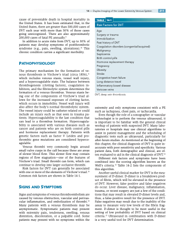
Risk factors for DVT can generally be associated with one or more of the elements of Virchow’s triad.12 Common risk factors are shown in Table 14-1.
SIGNS AND SYMPTOMS
Signs and symptoms of venous thromboembolism are caused by venous obstruction, vascular and perivas- cular inflammation, and embolization of thrombi.13 Many patients with a venous thrombosis may be asymptomatic. Symptomatic patients may present with extremity pain, tenderness, swelling, venous distention, discoloration, or a palpable cord. Some patients may present with no symptoms within the
TABLE 14-1
Risk Factors for DVT
Age
Surgery or trauma
Immobilization
Past history of DVT
Coagulation disorders (congenital/acquired) Malignancy
Septicemia
Birth control pills
Hormone replacement therapy
Pregnancy
Obesity
Stroke
Congestive heart failure
Long distance travel
Inflammatory bowel disease
Varicose veins
DVT, deep vein thrombosis.
extremity and only symptoms consistent with a PE such as tachypnea, chest pain, or tachycardia.
Even though the role of a sonographer or vascular technologist is to perform the venous ultrasound, it is important to be familiar with the general clinical workup of patients with suspected DVT. Many labo- ratories or hospitals may use clinical algorithms to assist in patient management and the scheduling of diagnostic tests such as ultrasound, particularly for after-hours studies. As mentioned at the beginning of this chapter, the clinical diagnosis of DVT is quite in- accurate with poor sensitivity and specificity. Various patient data, both demographic and clinical, are of- ten evaluated to aid in the clinical diagnosis of DVT.14
Different risk factors and symptoms have been combined into the scoring algorithm known as the Well’s criteria.15 Table 14-2 lists the components of this algorithm.
Another useful clinical marker for DVT is the mea- surement of D-dimer. D-dimer is a breakdown prod- uct of fibrin, which will be elevated in the presence of DVT. However, false positive and false negatives do occur. Liver disease, malignancy, inflammation, trauma, or recent surgery are just a few of the condi- tions that may result in elevated D-dimer levels and, thus, a false-positive result for the presence of DVT. False negatives may result due to the inability of the assay to measure very low levels of the fibrin frag- ment. D-dimer is thought to be most useful in the setting of low probability of DVT based on clinical criteria.15 Ultrasound in combination with D-dimer is a very useful tool for DVT determination.
