
Page 233 - Libro 2
P. 233
14 — Duplex Imaging of the Lower Extremity Venous System
213
institutions try to do without this transducer, but this is not the best course of action. This high-frequency transducer should have a small footprint (hockey stick–type transducers are very useful). This transducer (10-18 MHz) is used for superficial veins such as the saphenous in the leg and for most of the arm veins. This transducer is a must for detailed reflux studies or upper and lower extremity mapping.
• The third transducer needed for venous im- aging is the curved low-frequency transducer (2-5 MHz). This transducer is useful for the in- ferior vena cava (IVC) and iliac veins. It is also helpful in the heavy patient where some veins may lie deeper within the leg.
SCANNING TECHNIQUE
Recently, changes to the names of some of the veins in the leg were adopted that have unfortunately made things a bit more confusing. Some have been slow to adopt the new terms and others are not aware the changes have been made. In the following sections, both terms will be provided for those veins with new nomenclature. The protocol described here represents a summary of time-tested techniques for properly identifying the veins and evaluating them for a thrombus formation.16–19
Initial Examination Position
The lower extremity venous duplex examination be- gins at the groin crease. The common femoral vein (CFV) and common femoral artery (CFA) are located from a medial projection in a transverse plane. Mov- ing above this area and above the level of the in- guinal ligament, the CFV becomes the external iliac vein (EIV). Using the ultrasound transducer, gentle pressure is applied directly over the vein. Because the veins are under relatively low pressure, the walls of the vein will be seen to coapt or close together. The compression of the walls of the vein should be recorded. Remaining in a transverse plane, the com- pression and release technique is performed every 2 to 3 cm down the entire length of the leg, docu- menting the images at several locations along the leg. With each of the named vessels described as fol- lows, these compression maneuvers are performed and recorded. The smaller the “cuts” (or spacing be- tween compressions) used, the better. Spacing the cuts too far apart will allow for missing a smaller partial thrombus. After the entire vein has been im- aged in the transverse plane, the examiner can then reexamine the same vein in a longitudinal plane. This will add additional information and can be used to confirm findings observed in the transverse plane.
Doppler and color imaging can be performed in the longitudinal plane.
It cannot be overemphasized, however, that one cannot omit the transverse view and do only longitu- dinal imaging. Doing so will result in missing nonob- structive thrombi. It is very easy to roll off a vein while in the longitudinal plane; therefore, compressions should never be performed in a longitudinal view.
In addition to compression of the veins, spectral Doppler waveforms are usually recorded from the CFV and popliteal veins. Institutions vary at which specific veins Doppler waveforms should be ob- tained but generally it is at least at two levels. For a DVT examination, Doppler waveforms are examined strictly for qualitative features such as spontaneity, phasicity with respiration, augmentation with distal compression, and cessation of flow with proximal compression.
Common Femoral and Great Saphenous Veins
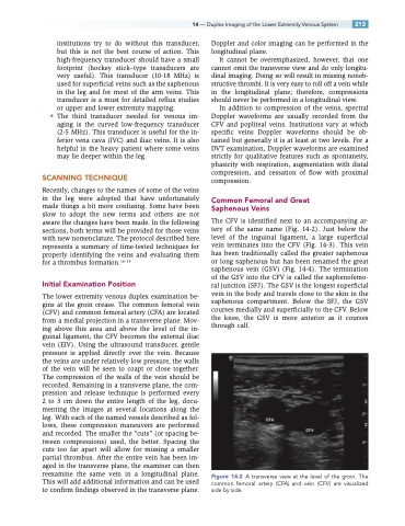
The CFV is identified next to an accompanying ar- tery of the same name (Fig. 14-2). Just below the level of the inguinal ligament, a large superficial vein terminates into the CFV (Fig. 14-3). This vein has been traditionally called the greater saphenous or long saphenous but has been renamed the great saphenous vein (GSV) (Fig. 14-4). The termination of the GSV into the CFV is called the saphenofemo- ral junction (SFJ). The GSV is the longest superficial vein in the body and travels close to the skin in the saphenous compartment. Below the SFJ, the GSV courses medially and superficially to the CFV. Below the knee, the GSV is more anterior as it courses through calf.
Figure 14-2 A transverse view at the level of the groin. The common femoral artery (CFA) and vein (CFV ) are visualized side by side.
